File:Boyden1931 fig10.jpg
Original file (800 × 800 pixels, file size: 86 KB, MIME type: image/jpeg)
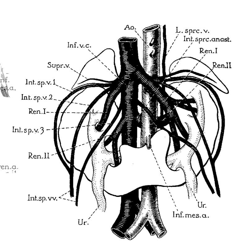
Fig. 10. Horseshoe kidney
Horseshoe kidney from a man about forty years of age (after Ssokolow, ’30). Note arrangement of multiple internal spermatic veins (Int.sp.v. 1, 2,3), the persistence and Well—marked separation of both embryonic renal veins (Ren. I and II), the persistence of an intersnpracardinal anastomosis behind the aorta (1'nt.s;m*c.anast.) connecting the right supracardinal (Inf.o.c.) with the persisting left supracardinal (L.spr1;.) and renal (Re'rL.II) veins.
Reference
Boyden EA. Description of a horseshoe kidney associated with left inferior vena cava and disc-shaped suprarenal glands, together with a note on the occurrence of horseshoe kidneys in human embryos. (1931) Anat. Rec. 51(2): 187-211.
Cite this page: Hill, M.A. (2024, May 16) Embryology Boyden1931 fig10.jpg. Retrieved from https://embryology.med.unsw.edu.au/embryology/index.php/File:Boyden1931_fig10.jpg
- © Dr Mark Hill 2024, UNSW Embryology ISBN: 978 0 7334 2609 4 - UNSW CRICOS Provider Code No. 00098G
File history
Click on a date/time to view the file as it appeared at that time.
| Date/Time | Thumbnail | Dimensions | User | Comment | |
|---|---|---|---|---|---|
| current | 10:35, 8 September 2017 | | 800 × 800 (86 KB) | Z8600021 (talk | contribs) |
You cannot overwrite this file.
File usage
The following 3 pages use this file: