File:Boyden1931 fig08.jpg
Original file (800 × 800 pixels, file size: 55 KB, MIME type: image/jpeg)
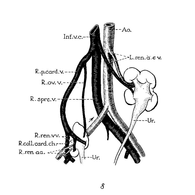
Fig. 8. Double ectopic, unrotated kidneys
Double ectopic, unrotated kidneys from a girl twenty-three years of age (after Melissinos, ’1l). Note position of diminutive right kidney on brim of pelvis, tl1e persistence of the right posterior cardinal vein (R.p.ea1'd.12.) and of the right collateral cardinal channel (I.’.coll.card.c7:..), the disappearance of the right common iliac vein (site indicated by arrow), and the origin of the right renal veins and arteries (R.ren.1m. and ac.) from the posterior cardinal vein and common iliac artery, respectively.
Reference
Boyden EA. Description of a horseshoe kidney associated with left inferior vena cava and disc-shaped suprarenal glands, together with a note on the occurrence of horseshoe kidneys in human embryos. (1931) Anat. Rec. 51(2): 187-211.
Cite this page: Hill, M.A. (2024, May 15) Embryology Boyden1931 fig08.jpg. Retrieved from https://embryology.med.unsw.edu.au/embryology/index.php/File:Boyden1931_fig08.jpg
- © Dr Mark Hill 2024, UNSW Embryology ISBN: 978 0 7334 2609 4 - UNSW CRICOS Provider Code No. 00098G
File history
Click on a date/time to view the file as it appeared at that time.
| Date/Time | Thumbnail | Dimensions | User | Comment | |
|---|---|---|---|---|---|
| current | 10:35, 8 September 2017 | | 800 × 800 (55 KB) | Z8600021 (talk | contribs) |
You cannot overwrite this file.
File usage
The following 2 pages use this file: