File:Bailey379-382.jpg
Original file (671 × 988 pixels, file size: 199 KB, MIME type: image/jpeg)
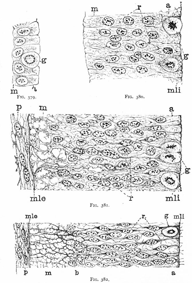
Fig. 379. From the neural tube of an embryo rabbit shortly before the closure of the tube
g, Germinal or dividing cell; w, peripheral zone, position of the later marginal layer. His.
Fig. 380. Pig of 5 mm, unflexed. Just after closure of the neural tube. Segment of a vertical section of the lateral wall of the tube, g, Germinal cells; m, beginning of marginal layer; mli, internal limiting membrane; r, radial columns of protoplasm. The resting nuclei lie in the inner or nuclear layer. Hardesty.
"With the further increase and development of the nervous elements (see p. 455) the radial arrangement of the spongioblasts noted above becomes more and more obliterated. As shown by Golgi preparations, in their migration from the lumen (Fig. 384) the spongioblasts lose their connection with the lumen, their peripheral processes become abbreviated and disappear, and they finally differentiate into the irregular branching neuroglia cells (Fig. 385). According to Hardesty, there is simply a general nucleated mass which changes form pari passu with changes in the enclosed . differentiating nervous elements, finally assuming shapes dependent upon the character of the spaces between the formed nervous elements. An exception to this is a layer of nucleated elements which remain next the lumen and form the ependyma cells which still send radial extensions into the wall of the neural tube (Figs. 383 and 384). These cells develop cilia projecting into the lumen.
A still later differentiation in the supporting elements of the tube is the appearance of neuroglia fibers a product of the spongioblastic protoplasm, but differing from it chemically (Fig. 385). The exact relation of these neuroglia fibers to the nucleated neuroglia cells in the adult is a matter of dispute.
- Text-Book of Embryology: Germ cells | Maturation | Fertilization | Amphioxus | Frog | Chick | Mammalian | External body form | Connective tissues and skeletal | Vascular | Muscular | Alimentary tube and organs | Respiratory | Coelom, Diaphragm and Mesenteries | Urogenital | Integumentary | Nervous System | Special Sense | Foetal Membranes | Teratogenesis | Gallery of All Figures
| Historic Disclaimer - information about historic embryology pages |
|---|
 |
Reference
Bailey FR. and Miller AM. Text-Book of Embryology (1921) New York: William Wood and Co.
Cite this page: Hill, M.A. (2024, May 8) Embryology Bailey379-382.jpg. Retrieved from https://embryology.med.unsw.edu.au/embryology/index.php/File:Bailey379-382.jpg
- © Dr Mark Hill 2024, UNSW Embryology ISBN: 978 0 7334 2609 4 - UNSW CRICOS Provider Code No. 00098G
File history
Click on a date/time to view the file as it appeared at that time.
| Date/Time | Thumbnail | Dimensions | User | Comment | |
|---|---|---|---|---|---|
| current | 00:18, 30 January 2011 | | 671 × 988 (199 KB) | S8600021 (talk | contribs) | ==Fig. 379. From the neural tube of an embryo rabbit shortly before the closure of the tube== g, Germinal or dividing cell; w, peripheral zone, position of the later marginal layer. His. '''Fig. 380. Pig of 5 mm, unflexed. Just after closure of the neu |
You cannot overwrite this file.
File usage
The following 3 pages use this file: