File:Bailey061.jpg

Original file (878 × 1,086 pixels, file size: 184 KB, MIME type: image/jpeg)
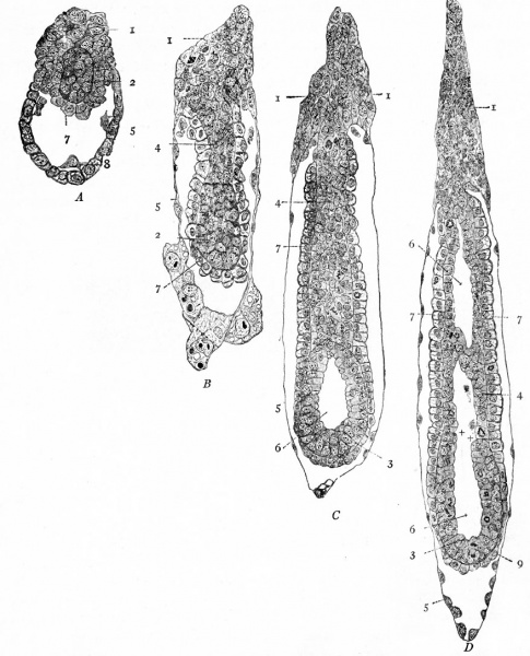
Fig. 61. Sections of blastocysts of the white rat, showing inversion (entypy) of the germ layers
Huber.
A, blastocyst 6 days and 14 hours after insemination.
B, blastocyst 8 days and 1 8 hours after insemination, with egg-cylinder cut longitudinally.
C, similar section of blastocyst 7 days and 22 hours after insemination (younger than B but further advanced in development), showing beginning of proamniotic cavity.
D, similar section of blastocyst 8 days after insemination (younger than B but further advanced in development), showing more advanced proamniotic cavity.
Legend
- Ectoplacental cone
- ectodermal node
- primary embryonic ectoderm
- extraembryonic ectoderm
- transitory ectoderm (original wall of blastocyst)
- proamniotic cavity
- visceral entoderm
- cells of parietal entoderm
- primary embryonic entoderm.
- Text-Book of Embryology: Germ cells | Maturation | Fertilization | Amphioxus | Frog | Chick | Mammalian | External body form | Connective tissues and skeletal | Vascular | Muscular | Alimentary tube and organs | Respiratory | Coelom, Diaphragm and Mesenteries | Urogenital | Integumentary | Nervous System | Special Sense | Foetal Membranes | Teratogenesis | Gallery of All Figures
| Historic Disclaimer - information about historic embryology pages |
|---|
 |
Reference
Bailey FR. and Miller AM. Text-Book of Embryology (1921) New York: William Wood and Co.
Cite this page: Hill, M.A. (2024, May 19) Embryology Bailey061.jpg. Retrieved from https://embryology.med.unsw.edu.au/embryology/index.php/File:Bailey061.jpg
- © Dr Mark Hill 2024, UNSW Embryology ISBN: 978 0 7334 2609 4 - UNSW CRICOS Provider Code No. 00098G
File history
Click on a date/time to view the file as it appeared at that time.
| Date/Time | Thumbnail | Dimensions | User | Comment | |
|---|---|---|---|---|---|
| current | 16:57, 17 January 2011 | | 878 × 1,086 (184 KB) | S8600021 (talk | contribs) | {{Template:Bailey 1921 Figures}} |
You cannot overwrite this file.
File usage
The following 3 pages use this file: