File:Bailey036.jpg
Original file (673 × 863 pixels, file size: 164 KB, MIME type: image/jpeg)
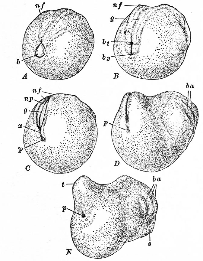
Fig. 36. Postero-lateral views of successive stages following gastrulation in the frog
Ziegler, from Kellicott.
A, blastopore in process of closing, neural folds slightly indicated;
B, gastrula slightly elongated, blastopore closed, neural groove and folds obvious;
C, anal portion of blastopore still visible at bottom of proctodaeum, neural folds closing dorsally;
D, neural folds nearly closed, branchial arches appearing, tail bud forming; E, neural folds fused, tail bud more conspicuous.
b, Blastopore containing yolk plug; bi, dorsal part of blastopore (rudiment of neurenteric canal) ; fe, ventral part of 'blastopore (rudiment of anus); ba, branchial arches; g, neural gro ve ; nf, neural folds; np, neural plate; p, proctodaeum, with anal portion of the blastopore at the bottom; s, oral sucker; t, tail bud; x, neural folds covering the blastopore thus establishing the neurenteric canal.
- Text-Book of Embryology: Germ cells | Maturation | Fertilization | Amphioxus | Frog | Chick | Mammalian | External body form | Connective tissues and skeletal | Vascular | Muscular | Alimentary tube and organs | Respiratory | Coelom, Diaphragm and Mesenteries | Urogenital | Integumentary | Nervous System | Special Sense | Foetal Membranes | Teratogenesis | Gallery of All Figures
| Historic Disclaimer - information about historic embryology pages |
|---|
 |
Reference
Bailey FR. and Miller AM. Text-Book of Embryology (1921) New York: William Wood and Co.
Cite this page: Hill, M.A. (2024, May 15) Embryology Bailey036.jpg. Retrieved from https://embryology.med.unsw.edu.au/embryology/index.php/File:Bailey036.jpg
- © Dr Mark Hill 2024, UNSW Embryology ISBN: 978 0 7334 2609 4 - UNSW CRICOS Provider Code No. 00098G
File history
Click on a date/time to view the file as it appeared at that time.
| Date/Time | Thumbnail | Dimensions | User | Comment | |
|---|---|---|---|---|---|
| current | 14:43, 17 January 2011 | | 673 × 863 (164 KB) | S8600021 (talk | contribs) | {{Template:Bailey 1921 Figures}} |
You cannot overwrite this file.
File usage
The following 3 pages use this file: