File:Anson1948 fig15.jpg

Original file (1,280 × 829 pixels, file size: 158 KB, MIME type: image/jpeg)
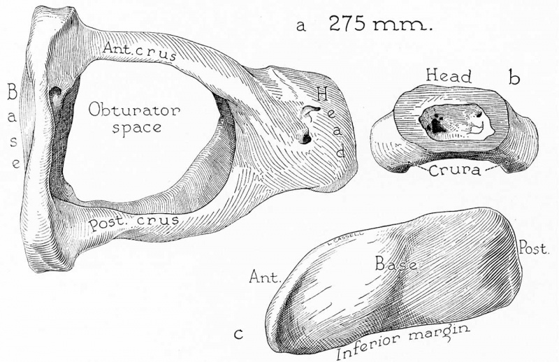
Fig. 15. Drawings of a reconstruction of the stapes in a 275 mm
(30 week) fetus (Wisconsin series 4) ; X 7: (a) the reconstruction entire, viewed from above (superior, or cranial, aspect); (b) the neck and adjacent portions of the crura, in lateral (tympanic) view; (c) the base of the ossicle seen in medial (vestibular) view.
The stapes has attained adult form; of the crura, now deeply channeled, the posterior is longer and larger (as); the base is flatter on the posterior than on the anterior margin (c) ; the head and neck are hollowed (b). In this fetal specimen, as in some adult ossicles, a ledge of bone partially closes the space of the neck toward the obturator foramen.
Reference
Anson BJ. and Cauldwell EW. Stapes, fissula ante fenestram and associated structures in man: V . From the fetus of 160 mm to term. (1948) 48(3): 263-300.
Cite this page: Hill, M.A. (2024, May 15) Embryology Anson1948 fig15.jpg. Retrieved from https://embryology.med.unsw.edu.au/embryology/index.php/File:Anson1948_fig15.jpg
- © Dr Mark Hill 2024, UNSW Embryology ISBN: 978 0 7334 2609 4 - UNSW CRICOS Provider Code No. 00098G
File history
Click on a date/time to view the file as it appeared at that time.
| Date/Time | Thumbnail | Dimensions | User | Comment | |
|---|---|---|---|---|---|
| current | 21:34, 16 October 2017 | | 1,280 × 829 (158 KB) | Z8600021 (talk | contribs) | |
| 21:32, 16 October 2017 | 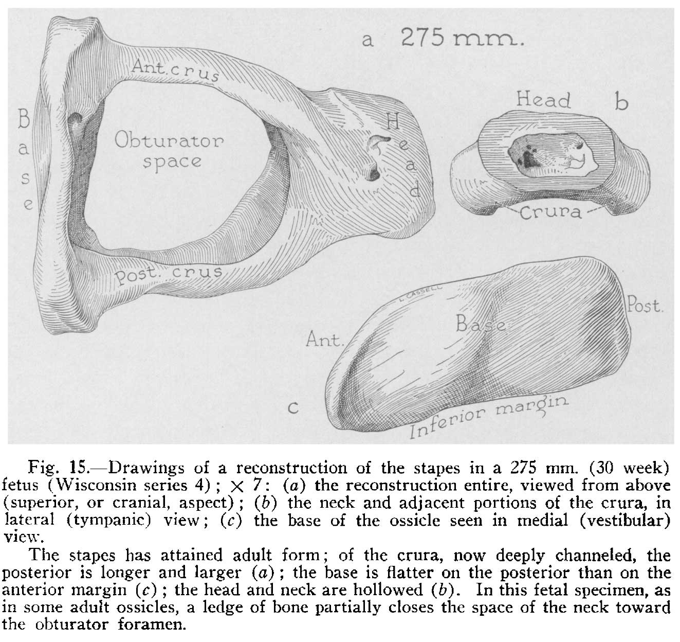 | 1,323 × 1,248 (211 KB) | Z8600021 (talk | contribs) | ===Reference=== {{Ref-Anson1948}} {{Footer}} Category:Middle EarCategory:Historic EmbryologyCategory:1940's |
You cannot overwrite this file.
File usage
The following 2 pages use this file: