Category:Testis
From Embryology
This Embryology category shows media and pages related to the male gonad, the testis.
| Male | Y | SRY | testis | spermatozoa | ductus deferens | penis | prostate | Category:Male |
Pages in category 'Testis'
The following 86 pages are in this category, out of 86 total.
A
B
M
P
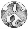
- Paper - A morphological study of testicular descent
- Paper - A study of the function of the epididymis 1 (1929)
- Paper - Development and transition of the testis, normal and abnormal 1
- Paper - Development and transition of the testis, normal and abnormal 2
- Paper - Development and transition of the testis, normal and abnormal 3
- Paper - Development and transition of the testis, normal and abnormal 4
- Paper - Development and vascularization of the testis (1906)
- Paper - Has a persistence of the Müllerian ducts any relation to the conditions of cryptorchidism?
- Paper - Studies on the fine structure of the mammalian testis 1
- Paper - The Embryonic Development of the Interstitial Cells of Leydig (1904)
- Paper - The histology of the retained testis in the human subject at different ages, and its comparison with the scrotal testis (1929)
- Paper - The inguinal canal in the foetus and new-born (1944)
- Paper - The morphology of the seminiferous tubules of Mammalia (1913)
- Template:Peritubular myoid cell
- Template:Persistent Müllerian duct syndrome
- Template:Primary spermatocyte
R
- Template:Ref-Allen1904
- Template:Ref-BascomOsterud1925
- Template:Ref-Bremer1911
- Template:Ref-BurgosFawcett1955
- Template:Ref-Cooper1929
- Template:Ref-Crew1922
- Template:Ref-Hill1906
- Template:Ref-Lockwood1887a
- Template:Ref-Lockwood1887b
- Template:Ref-Lockwood1888a
- Template:Ref-Lockwood1888b
- Template:Ref-PelliniemiNiemi1969
- Template:Ref-RowlandsBrambell1932
- Template:Ref-Young1929
- Template:Ref-Young1961
S
T
Media in category 'Testis'
The following 169 files are in this category, out of 169 total.
 Adrenal and gonad early development.jpg 700 × 397; 50 KB
 Adrenal and gonad steroidogenic factor 1 expression.jpg 1,000 × 636; 88 KB
 Bailey309.jpg 594 × 592; 58 KB
 Bailey327.jpg 872 × 567; 89 KB
 Bailey332.jpg 637 × 356; 53 KB
 Bailey337.jpg 790 × 573; 74 KB
 Bailey338.jpg 940 × 473; 87 KB
 Bailey341.jpg 832 × 675; 69 KB
 BurgosFawcett1955 fig11.jpg 1,453 × 2,015; 528 KB
 BurgosFawcett1955 fig13.jpg 1,460 × 2,049; 501 KB
 BurgosFawcett1955 fig14.jpg 1,456 × 1,965; 381 KB
 BurgosFawcett1955 text-fig01.jpg 1,280 × 1,137; 143 KB
 Corner1920 fig01.jpg 1,000 × 606; 159 KB
 Cryptorchidism.jpg 600 × 390; 35 KB
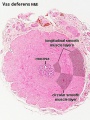 Ductus deferens 01.jpg 400 × 533; 76 KB
 Ductus deferens 02.jpg 400 × 533; 80 KB
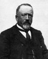 Enrico Sertoli.jpg 650 × 800; 68 KB
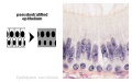 Epididymis histology 01.jpg 600 × 375; 20 KB
 Epididymis histology 02.jpg 400 × 534; 71 KB
 Epididymis histology 03.jpg 400 × 533; 68 KB
 Fetal gonad retinoid receptor expression 01.jpg 1,004 × 1,000; 226 KB
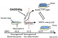 Gadd45g and sex determination model.jpg 909 × 600; 62 KB
 Germ cell tumor 02.jpg 800 × 599; 168 KB
 Gray1114.jpg 450 × 471; 47 KB
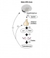 HPG male axis.jpg 600 × 700; 36 KB
 Human fetal gonad retinoid receptor expression.jpg 1,004 × 1,000; 447 KB
 Keibel Mall 008.jpg 1,200 × 515; 161 KB
 Keibel Mall 2 633.jpg 1,000 × 681; 74 KB
 Keibel Mall 2 635.jpg 1,000 × 1,073; 223 KB
 Keibel Mall 2 636.jpg 1,200 × 738; 119 KB
 Keibel Mall 2 658a.jpg 1,127 × 1,200; 103 KB
 Keibel Mall 2 658b.jpg 895 × 1,200; 98 KB
 Keith1902 fig102.jpg 800 × 605; 68 KB
 Keith1902 fig104.jpg 800 × 601; 77 KB
 Kollmann013.jpg 732 × 626; 95 KB
 Kollmann445.jpg 776 × 738; 112 KB
 Kollmann446.jpg 723 × 414; 38 KB
 Kollmann447.jpg 564 × 556; 42 KB
 Kollmann448.jpg 643 × 576; 43 KB
 Kollmann449.jpg 613 × 617; 44 KB
 Kollmann458.jpg 1,000 × 520; 120 KB
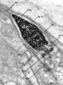
 Leydig cell PMID13693345 EM02.jpg 1,359 × 957; 341 KB
 Leydig cell PMID13693345 EM03.jpg 1,359 × 957; 325 KB
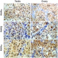
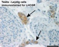 Leydig cells stained for LHCGR1.jpg 404 × 322; 16 KB
 Lockwood1887b fig23.jpg 500 × 423; 41 KB
 Lockwood1887b fig24.jpg 600 × 463; 82 KB
 Lockwood1887b fig25.jpg 715 × 1,000; 125 KB
 Lockwood1887b fig26.jpg 500 × 266; 38 KB
 Lockwood1887b fig27.jpg 500 × 378; 46 KB
 Lockwood1887b fig28.jpg 600 × 592; 43 KB
 Lockwood1887b fig29.jpg 800 × 656; 121 KB
 Lockwood1887b fig30.jpg 800 × 619; 69 KB
 Lockwood1887b fig31.jpg 800 × 576; 49 KB
 Lockwood1887b fig32.jpg 500 × 329; 55 KB
 Lockwood1887b fig33.jpg 600 × 617; 101 KB
 Lockwood1887b fig34.jpg 408 × 800; 43 KB
 Lockwood1887b fig35.jpg 800 × 600; 117 KB
 Lockwood1887b fig36.jpg 600 × 828; 53 KB
 Lockwood1887b fig37.jpg 800 × 739; 102 KB
 Lockwood1887b fig38.jpg 800 × 688; 65 KB
 Lockwood1887b fig39.jpg 800 × 1,102; 93 KB
 Lockwood1887b fig40.jpg 774 × 1,000; 92 KB
 Lockwood1887b fig41.jpg 800 × 547; 129 KB
 Lockwood1887b plate02.jpg 2,998 × 2,272; 1.21 MB
 Lockwood1888a fig47.jpg 1,000 × 712; 279 KB
 Lockwood1888a fig50.jpg 959 × 652; 85 KB
 Lockwood1888a plate07.jpg 1,280 × 985; 285 KB
 Lockwood1888b fig49.jpg 800 × 496; 69 KB
 Lockwood1888b fig51.jpg 800 × 725; 88 KB
 Lockwood1888b fig52.jpg 800 × 824; 60 KB
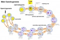 Male gametogenesis.jpg 1,000 × 666; 121 KB
 Male puberty testicular volume graph.jpg 1,140 × 826; 126 KB
 Model male androsterone synthesis.jpg 740 × 518; 92 KB
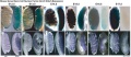
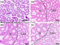 Mouse epididymis development 01.jpg 1,200 × 909; 486 KB
 Mouse epididymis development 02.jpg 600 × 451; 138 KB
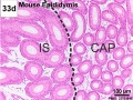 Mouse epididymis development 03.jpg 600 × 451; 119 KB
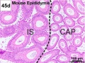 Mouse epididymis development 04.jpg 600 × 451; 120 KB
 Mouse epididymis development 05.jpg 600 × 451; 112 KB
 Mouse gonad development timeline.jpg 1,200 × 697; 98 KB
 Mouse gonad Gcnf expression 01.jpg 1,947 × 843; 304 KB
 Mouse gonad Gcnf expression E12.5.jpg 331 × 785; 68 KB
 Mouse gonad Gcnf expression E13.5.jpg 332 × 784; 60 KB
 Mouse gonad Gcnf expression E14.5.jpg 334 × 784; 60 KB
 Mouse gonad Gcnf expression E15.5.jpg 338 × 782; 53 KB
 Mouse gonad Gcnf expression E16.5.jpg 325 × 786; 40 KB
 Mouse gonad Gcnf expression E17.5.jpg 328 × 786; 44 KB
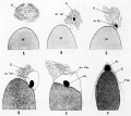
 Mouse gonad sex determination 01.jpg 600 × 600; 81 KB
 Mouse sex determination genes 01.jpg 1,280 × 923; 73 KB
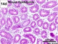
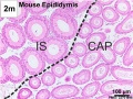
 Mouse- epididymis histology.jpg 751 × 383; 82 KB
 Mouse- gonadal supporting cell development.jpg 1,000 × 588; 74 KB
 Nelsen1953 fig002.jpg 1,200 × 1,037; 249 KB
 Nelsen1953 fig004.jpg 1,200 × 960; 200 KB
 Nelsen1953 fig005.jpg 1,200 × 709; 102 KB
 Nelsen1953 fig006.jpg 1,200 × 732; 224 KB
 Nelsen1953 fig007.jpg 1,200 × 1,311; 343 KB
 Orchidometer.jpg 361 × 225; 14 KB
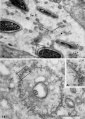
 Rat blood–testis barrier 01.jpg 1,002 × 1,599; 221 KB
 Rat blood–testis barrier 02.jpg 1,002 × 853; 125 KB
 Rat- immortal germ cells are spermatogonial stem cells.jpg 459 × 1,000; 72 KB
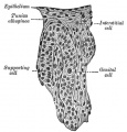
 Seminiferous tubule cartoon.jpg 800 × 544; 92 KB
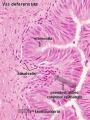
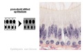
 Seminiferous-tubule-HEx40.jpg 400 × 500; 59 KB
 Simkins1928 plate01.jpg 1,574 × 2,003; 237 KB
 Simkins1928 plate02.jpg 1,464 × 2,126; 223 KB
 Simkins1928 plate03.jpg 1,551 × 2,086; 293 KB
 Simkins1928 plate04.jpg 1,543 × 2,026; 219 KB
 Simkins1928 plate05.jpg 1,281 × 2,111; 154 KB
 Simkins1928 plate06.jpg 1,587 × 2,088; 277 KB
 Simkins1928 plate07.jpg 1,540 × 2,096; 239 KB
 Simkins1928 plate08.jpg 1,558 × 1,797; 220 KB
 Simkins1928 plate09.jpg 1,548 × 2,096; 263 KB
 Simkins1928 plate10.jpg 1,565 × 1,386; 140 KB
 Spermatogenesis cartoon 01.jpg 1,064 × 759; 142 KB
 Spermatozoa histology 001.jpg 1,280 × 1,024; 366 KB
 Spermatozoa histology 002.jpg 1,280 × 1,024; 246 KB
 Spermatozoa histology 003.jpg 1,280 × 1,024; 166 KB
 Stage 22 image 188.jpg 1,000 × 665; 112 KB
 Stage 22 image 191.jpg 1,000 × 653; 100 KB
 Stage 22 image 194.jpg 1,000 × 671; 210 KB
 Stage 22 image 195.jpg 1,000 × 657; 265 KB
 Stage 22 image 201.jpg 1,200 × 754; 324 KB
 Stage 22 image 202.jpg 1,455 × 920; 617 KB
 Stage 22 image 301.jpg 1,200 × 754; 329 KB
 Stage 22 image 302.jpg 1,455 × 920; 625 KB
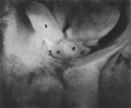
 Suprascrotal testis.jpg 1,000 × 751; 126 KB
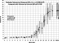 Testicular volume graph.jpg 554 × 405; 57 KB
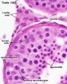
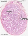
 Testis histology 001.jpg 1,280 × 1,024; 574 KB
 Testis histology 002.jpg 1,280 × 1,024; 599 KB
 Testis histology 003.jpg 1,280 × 1,024; 183 KB
 Testis histology 004.jpg 1,280 × 1,024; 396 KB
 Testis histology 005.jpg 1,280 × 1,024; 266 KB
 Testis histology 006.jpg 1,280 × 1,024; 251 KB
 Testis histology 007.jpg 1,280 × 1,024; 256 KB
 Testis histology 008.jpg 1,280 × 1,024; 454 KB
 Testis histology 009.jpg 1,280 × 1,024; 339 KB
 Testis histology 010.jpg 1,280 × 1,024; 422 KB
 Testis histology 011.jpg 1,280 × 1,024; 245 KB
 Testis histology 012.jpg 1,280 × 1,024; 266 KB
 Testis histology 013.jpg 1,280 × 1,024; 418 KB
 Testis histology 014.jpg 1,280 × 1,024; 352 KB
 Testis histology 015.jpg 1,280 × 1,024; 281 KB
 Testis histology 016.jpg 1,280 × 1,024; 322 KB
 Testis histology 017.jpg 1,280 × 1,024; 283 KB
 Testis histology 018.jpg 1,280 × 1,024; 350 KB
 Testis histology 019.jpg 1,280 × 1,024; 239 KB
 Testis histology 02.jpg 246 × 481; 49 KB
 Testis histology 020.jpg 1,300 × 685; 334 KB
 Testis histology 021.jpg 1,200 × 962; 312 KB
 Testis histology 022.jpg 1,229 × 966; 311 KB
 Testis histology 023.jpg 600 × 375; 35 KB
 Testis histology 1.jpg 400 × 500; 113 KB
 Testis histology 2.jpg 400 × 500; 32 KB
 Testis histology.jpg 400 × 500; 54 KB
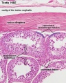
 Testis, young H&E reproductive system, male, convoluted seminiferous tubules x10.jpg 1,280 × 1,024; 396 KB
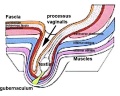 Testis-descent end.jpg 600 × 480; 33 KB
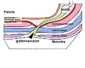 Testis-descent start.jpg 600 × 480; 26 KB
 Wyndham1943 fig01.jpg 800 × 660; 70 KB
 Wyndham1943 fig02.jpg 733 × 768; 172 KB
 Wyndham1943 fig03.jpg 756 × 754; 189 KB
 Wyndham1943 fig04.jpg 742 × 751; 195 KB
 Wyndham1943 fig05.jpg 632 × 741; 120 KB
 Wyndham1943 fig06.jpg 641 × 814; 166 KB
 Wyndham1943 fig07.jpg 779 × 650; 127 KB
 Wyndham1943 fig08.jpg 886 × 638; 171 KB
 Wyndham1943 fig09.jpg 636 × 1,000; 198 KB
 Wyndham1943 fig10.jpg 605 × 1,000; 183 KB
 Wyndham1943 fig11.jpg 1,000 × 447; 96 KB
 Wyndham1943 plate01.jpg 2,004 × 2,469; 719 KB
 Wyndham1943 plate02.jpg 2,004 × 2,471; 990 KB
 Wyndham1943 plate03.jpg 2,004 × 2,464; 1.08 MB


