Category:Student Image
From Embryology
Content in this category relates to images added to the site by students.
As shown by the text below:
Note - This image was originally uploaded as part of a student project and may contain inaccuracies in either description or acknowledgements.
--MarkHill 13:50, 4 February 2011 (EST) This category was added in February 2011 and may not appear on images uploaded before this time.
Pages in category 'Student Image'
The following 11 pages are in this category, out of 11 total.
Media in category 'Student Image'
The following 200 files are in this category, out of 650 total.
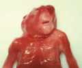
(previous page) (next page) 1 week year old newborn girl with cleft lip and palate.png 1,984 × 2,964; 2.67 MB
 1hroldrabbit.jpg 800 × 532; 102 KB
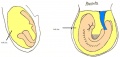
 2 day old embryo diagram.jpeg 505 × 243; 18 KB
 200px-Overlapping fingers.JPG 200 × 150; 9 KB
 220px-ClubbingCF.JPG 220 × 165; 8 KB
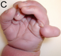 220px-Patauhand.PNG 220 × 200; 69 KB
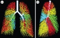 3D model of the air way tree.jpg 1,280 × 819; 215 KB
 3D Organoids.jpg 1,600 × 708; 1.11 MB
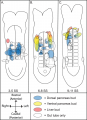 3–11 SS caudal foregut endoderm.png 1,347 × 1,855; 315 KB
 5months-gestation-retina.jpg 527 × 750; 173 KB
 77254175 embryo repair 624 method 2.gif 624 × 420; 35 KB
 77260486 cell structure 304.gif 304 × 228; 19 KB
 77266645 embryo repair 624 method 1.gif 624 × 407; 32 KB
 800px-Knockout Mice5006-300.jpg 800 × 521; 71 KB
 Abdominal Paracentesis.png 1,024 × 1,024; 825 KB
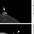 Abnormal formation of fertilisation cones .jpg 256 × 256; 5 KB
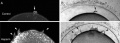 Abnormal Formation of Fertilisation Cones.jpg 638 × 228; 79 KB
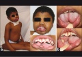 Abnormal of tongue.jpg 743 × 520; 82 KB
 Abnormal pectoral fins are formed in Notch signalling disrupted larvae.jpg 709 × 1,365; 222 KB
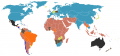 AbortionLawMap.png 800 × 370; 139 KB
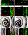
 Absence of CSFR1 Impacts Normal Development of Brain Architecture.jpg 596 × 595; 111 KB
 ACGH tracing after trophectoderm biopsy.jpeg 774 × 828; 132 KB
 ACGH.jpg 2,409 × 3,436; 474 KB
 Active parathyroid hormone assay cartoon.png 2,631 × 1,981; 822 KB
 Adaptation Cerebellum.png 2,918 × 1,828; 2.71 MB
 Addiction Cerebellum.png 1,352 × 488; 928 KB
 Adrenal gland.png 313 × 523; 90 KB
 Adrenal Medulla Developmental Timeline.jpg 4,160 × 1,658; 1.02 MB
 Ago2 in Mammalian Gastrulation.png 3,326 × 3,486; 7.49 MB
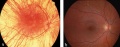 Albino fundus.jpg 333 × 131; 21 KB
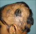 Alopecia Areata.jpg 571 × 538; 99 KB
 Amnion fold development in chicken embryos.jpg 786 × 443; 94 KB
 Amphioxus development.gif 778 × 884; 284 KB
 An overview of the process of fertilisation in mutant C. elegans.jpeg 1,325 × 2,800; 1.15 MB
 Anatomical diagram of testes.jpg 647 × 834; 237 KB
 Anatomical-Lobes-of-the-Cerebellum.jpg 994 × 468; 102 KB
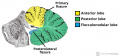
 Anatomy cerebral cortex.png 994 × 695; 713 KB
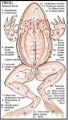 ANATOMY OF FROG.jpg 252 × 445; 37 KB
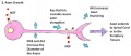
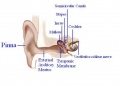 Anatomy of the Ear.JPG 566 × 405; 23 KB
 Anencephaly.png 652 × 544; 343 KB
 Angelman Syndrome Prenatal Diagnosis.jpg 720 × 540; 69 KB
 Annular pancreas.jpg 600 × 265; 16 KB
 Anterior Pituitary Hormones.jpg 640 × 480; 74 KB
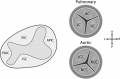
 Aortic valve disease.jpg 434 × 489; 241 KB
 Appearance of cornea due to CHED.png 235 × 233; 85 KB
 Aquaporins.png 1,927 × 2,863; 2.05 MB
 Arachnoid cyst with hydrocephalus.jpg 638 × 523; 72 KB
 Aristotle-eye.jpg 3,000 × 2,134; 351 KB
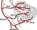 Arteries of the cerebellum.jpeg 329 × 267; 51 KB
 Ascension of the Kidneys.jpg 3,264 × 1,952; 936 KB
 Aspiration of a Blastomere.jpeg 600 × 487; 66 KB
 Aspiration of the Blastocoel Fluid.jpeg 600 × 462; 84 KB
 Astrocyte-vegf-deletion.JPG 495 × 600; 75 KB
 Atoh1 hair cell loss.png 2,074 × 2,521; 4.47 MB
 Atoh1 model.png 2,074 × 2,436; 3.5 MB
 Atrial Septal Defect (ASD).png 505 × 589; 337 KB
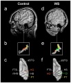 Auditory Cortex Location - Comparison Between Control Subject and WS Subject.jpg 1,084 × 1,261; 234 KB
 Axon growth.JPG 936 × 403; 40 KB
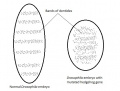 Bands of denticles in normal and Hh mutant Drosophila embryo.jpeg 491 × 374; 51 KB
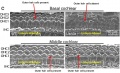 Basal Cochlear Outer Hair Cells.jpg 935 × 572; 114 KB
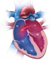
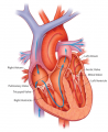 Basic anatomy of the heart.png 375 × 460; 203 KB
 Bilateral Stenosis of Internal Auditory canal.jpg 642 × 337; 125 KB
 Biopsy of Morula Stage Embryo.png 1,186 × 626; 705 KB
 Bone signalling pathway.gif 200 × 122; 12 KB
 Brachyury expression in 7.5dpc CD1 mouse embryos.jpg 386 × 313; 14 KB
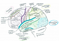
 Brain anatomy diagram .pdf ; 547 KB
 Brain Development 2.png 1,490 × 986; 2.12 MB
 Brain Development.png 1,390 × 842; 1.92 MB
 Brain sectin showing cortex.jpg 475 × 380; 25 KB
 Branching.jpg 634 × 407; 70 KB
 Canonical and non-canonical signalling TGF beta pathways.png 546 × 419; 254 KB
 Canonical Wnt pathway.jpg 500 × 375; 42 KB
 Capillary haemangioma.jpg 741 × 531; 116 KB
 Cardiac Development Overview .jpg 635 × 333; 51 KB
 Cardiac gene induction rates.png 4,103 × 2,460; 69 KB
 Cardiac melanocyte in an embryonic mouse pup.png 1,424 × 992; 427 KB
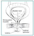 Cartoon of female urethra and bladder.jpg 250 × 266; 22 KB
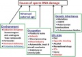 Causes of Increased DNA Damage.jpg 539 × 376; 54 KB
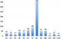 Cell number distribution for human embryos on day 3.png 1,542 × 990; 87 KB
 Cell signalling in cerebellum development.jpg 758 × 389; 230 KB
 Celsus-eye.jpg 1,700 × 1,722; 204 KB
 Cerebellum anatomical subdivisions.png 914 × 637; 67 KB
 Cerebellum Dystonia.png 2,094 × 490; 379 KB
 Cerebellum Layers and Cell Types.png 945 × 644; 103 KB
 Cerebellum Structure.jpg 1,280 × 392; 87 KB
 CffDNA from apoptotic trophoblasts.jpg 3,028 × 1,696; 1,019 KB
 Chart of mouse development 1.JPG 663 × 343; 32 KB
 Chart of mouse development 2.JPG 663 × 347; 31 KB
 Chemotherapy Side Effects 1.jpg 670 × 694; 105 KB
 Chemotherapy Treatment.jpeg 700 × 365; 58 KB
 Chemotherapy via Vein Infusion.jpg 475 × 404; 58 KB
 Chestxrayfallot.jpg 630 × 630; 136 KB
 Chew et al 2012 Figure 2 Tammar wallaby limb formation.jpg 3,531 × 1,594; 379 KB
 ChiariMalformation.jpg 358 × 434; 36 KB
 Chicken brain gene expression.jpg 1,200 × 530; 138 KB
 Chicken embryo E-cad and P-cad gastrulation.png 320 × 408; 253 KB
 Choanal atresia computed tomography 01.jpg 598 × 477; 35 KB
 Chromosome 15 Deletion of 15q11.2 to 15q13.jpg 222 × 548; 18 KB
 Cleavage - user.png 740 × 169; 177 KB
 Cleavage stage embryo.png 3,439 × 3,444; 7.13 MB
 Cleftp.jpg 756 × 1,044; 114 KB
 Clinical appearance of anophthalmia and microphthalmia.png 197 × 336; 98 KB
 Clinical examination TOF.png 479 × 350; 347 KB
 Clitoris.jpg 502 × 599; 102 KB
 Cloacal extrophy.jpg 708 × 286; 79 KB
 Cloacal partition completed.png 462 × 400; 60 KB
 CNS active.jpg 966 × 640; 165 KB
 CNS passive.jpg 960 × 378; 125 KB
 Cochlea stereocilia bundle.jpg 482 × 342; 31 KB
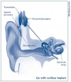 Cochlear Implant.jpg 428 × 492; 21 KB
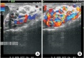 Color Doppler ultrasonography of varicocele.jpeg 666 × 461; 103 KB
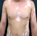 Common Vitiligo.jpg 600 × 583; 87 KB
 Components and structure of Spermatozoa.jpeg 600 × 783; 67 KB
 Corneal endothelial cell N-cadherin stained.png 320 × 265; 109 KB
 Cortex-group project.jpg 475 × 380; 25 KB
 Cortical growth rate patterns (redrawn diagram).JPG 631 × 366; 37 KB
 Cortical plate development.jpg 1,600 × 2,648; 4.02 MB
 Cortical thickness in Bmp7 knockouts.png 1,774 × 806; 1.93 MB
 Corticogenesis of mouse and humans.jpeg 1,024 × 770; 66 KB
 Corticogenesis.png 1,206 × 792; 211 KB
 CPAM.jpg 600 × 600; 47 KB
 CPAMXCT.jpg 1,559 × 838; 157 KB
 Cross section of genital tubercle male.jpg 576 × 886; 84 KB
 Cryopreservation of Porcine Embryos.jpeg 800 × 603; 111 KB
 CST Is Required for Vertebrate Heart Development.PNG 635 × 632; 570 KB
 Cushing's syndrome.jpg 450 × 592; 62 KB
 CVS Table.jpg 559 × 639; 94 KB
 DandyWalkerMalformation.jpeg 600 × 452; 63 KB
 Day 10 Closure of posterior neuropore, hind limb bud and tail bud.JPG 1,089 × 555; 76 KB
 Day 10.5 Deep Lens Indentation.JPG 514 × 483; 28 KB
 Day 11 Closure of lens vesicle.JPG 560 × 528; 27 KB
 Day 11.5 Lens Vesicle completely separated from surface.JPG 511 × 484; 29 KB
 Day 12 Earliest signs of fingers.JPG 510 × 466; 25 KB
 Day 13 Anterior Footplate Indented, marked pinna.JPG 437 × 470; 18 KB
 Day 14 Fingers Separate.JPG 517 × 541; 28 KB
 Day 15 toes separate.JPG 425 × 537; 22 KB
 Day 16 Reposition of umbilical hernia.JPG 640 × 532; 34 KB
 Day 17 fingers and toes joined together.JPG 425 × 554; 20 KB
 Day 18 long whiskers.JPG 462 × 514; 21 KB
 Day 19 Newborn Mouse.JPG 448 × 518; 18 KB
 Day 6 Differentiation of Egg Cylinder.JPG 425 × 498; 14 KB
 Day 6.5 Advanced Endometrial Reaction.JPG 425 × 556; 17 KB
 Day 7 Amnion.JPG 425 × 559; 18 KB
 Day 7.5 Neural plate, presomite stage.JPG 425 × 537; 23 KB
 Day 8 First Somites.JPG 1,087 × 590; 82 KB
 Day 8.5 Turning of embryo.JPG 1,173 × 557; 65 KB
 Day 9 Formation and closure of anterior neuropore.JPG 945 × 558; 54 KB
 Day 9.5 Formation of posterior neuropore and forelimb bud.JPG 752 × 568; 37 KB
 Deer mice oocytes at various stages of development in vitro.jpg 686 × 338; 56 KB
 Degenerated mouse embryos.jpeg 600 × 313; 47 KB
 Degradation of oocytes by sperm proteasomes.png 320 × 399; 125 KB
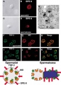
 Detection and Localisation of HPV in Sperms.png 600 × 238; 288 KB
 Development of Gonadotropin Preparations.jpeg 697 × 440; 76 KB
 Development of hypothalamus.jpg 1,600 × 468; 230 KB
 Development of nuclear transfer embryos.jpeg 689 × 482; 85 KB
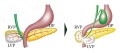
 Development of the pancreas during fetal development.jpg 600 × 332; 19 KB
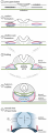
 Development of the Semilunar Valves.jpg 1,050 × 695; 105 KB
 Development of Trilaminar Embryonic Disc .png 1,921 × 5,223; 1.24 MB
 DevelopmentEye-PMC43029243.jpg 654 × 594; 142 KB
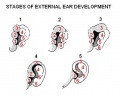 Devt of external ear.JPG 399 × 320; 20 KB
 Different forms of cleft palate.png 580 × 250; 259 KB
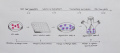
 Different Stages of Embryo Development.jpeg 567 × 279; 36 KB
 DNA-biological target of radiation.jpg 542 × 245; 38 KB
 Doctor performing an ECG on patient.png 539 × 334; 304 KB
 Dorsal Root Ganglia Adult.jpg 544 × 424; 160 KB
 Double tetrasomy 18 mosaicism.jpg 600 × 447; 78 KB
 Drawing of Tongue.png 800 × 600; 51 KB
 Duplicated ureter .jpg 1,050 × 539; 159 KB
 Dynamic Localization of Two Membrane Proteins for Fertilization.jpg 580 × 805; 88 KB
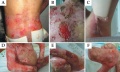 Dystrophic Epidermolysis Bullosa lesions.jpg 771 × 462; 115 KB
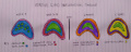
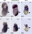
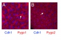 E18.5 developing kidney expressing Pygo1 and Pygo2.jpg 1,200 × 787; 260 KB
 Ectoderm Specification of Human Embryonic Stem Cells.png 2,209 × 2,042; 3.67 MB
 Effect of DMSO on asymmetric cell division in mouse oocytes.jpg 600 × 1,079; 126 KB
 Effect-of-vegf-on-retinal-vasculature.JPG 374 × 600; 50 KB
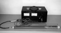 Electroejaculator.jpeg 637 × 350; 60 KB
 Electron micrographs of granulosa cells (GC).jpg 600 × 431; 99 KB
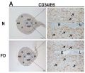 Embryo and Uterus stained with CD34 at Embryonic day 6.png 1,024 × 880; 876 KB
 Embryo development from pro-nuclei stage to blastocyst stage.jpeg 600 × 915; 42 KB
 Embryo Lung 1.png 730 × 853; 452 KB
 Embryonic dorsal root ganglia in mouse.jpg 765 × 599; 84 KB
 Embryonic Trachea.png 468 × 326; 205 KB
 Embryos during late blastula phase and early gastrulation.jpg 567 × 285; 64 KB
 Enamel Hypoplasia Due to Maternal Toxemia.jpg 700 × 466; 38 KB
 Endometrium structure.JPG 1,777 × 819; 99 KB
 Enlarged Vestibular aqueduct.jpg 495 × 289; 86 KB
 Eosin staining of mouse embryonic skin.png 629 × 251; 283 KB
 Epigenetic factors Influencing Human Development.jpg 567 × 393; 78 KB
 Ethanol fetal neural.jpg 827 × 597; 196 KB
 Examples of congenital defects in Apob and Lp mutant mice.gif 472 × 167; 43 KB
 Expression of BMP protein 4 .jpg 600 × 474; 148 KB
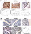
 Expression of CD82 in human placental villi and cell lines.JPG 612 × 432; 75 KB
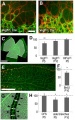
 Expression of Endometrial CD98 in implantation.jpg 996 × 1,090; 174 KB
 Expression of Endometrial CD98 in implantation.png 992 × 1,085; 5.66 MB
 External genitalia current model.jpg 638 × 899; 191 KB


.png)


.JPG)


.jpg)


