Category:Renal
From Embryology
This Embryology category shows media and pages related to renal system development. This includes the kidney, ureters, urinary bladder and urethra.
Subcategories
This category has the following 4 subcategories, out of 4 total.
Pages in category 'Renal'
The following 200 pages are in this category, out of 205 total.
(previous page) (next page)2
A
B
- Template:Bladder
- Template:Bladder exstrophy
- Book - A Laboratory Manual and Text-book of Embryology 8
- Book - Contributions to Embryology Carnegie Institution No.131
- Book - Manual of Human Embryology 19
- Book - Manual of Human Embryology 19-1
- Book - Manual of Human Embryology 19-2
- Book - Manual of Human Embryology 19-3
- Book - Manual of Human Embryology 19-4
- Book - Quain's Embryology 10
- Book - Text-Book of Embryology 15
C
E
H
I
- Template:ICD Renal (Q60-Q64)
- Template:ICD-11 Renal Agenesis or Dysgenesis anomalies collapse table
- Template:ICD-11 Renal Agenesis or Dysgenesis anomalies table
- Template:ICD-11 Renal anomalies header table
- Template:ICD-11 Ureter and Urethra anomalies collapse table
- Template:ICD-11 Ureter and Urethra anomalies table
- Template:Intermediate mesoderm
M
P
- Paper - A case of congenital absence of the left kidney and ureter (1915)
- Paper - A case of congenital cystic kidney, associated with several other abnormalities (1925)
- Paper - A case of duplication of the ureters (1915)
- Paper - A note on the post-natal growth of the kidney, thyroid gland and liver (1924)
- Paper - A three weeks human embryo with especial reference to the brain and nephric system (1905)
- Paper - An experimental study of the development of the amphibian cloaca (1940)
- Paper - Anomalies of the genito-urinary tract
- Paper - Congenital absence of the kidney
- Paper - Congenital cystic dilatation of the renal collecting tubules (1951)
- Paper - Congenital malformations of the ureters (1912)
- Paper - Congenital renal anomalies: with special reference to horseshoe kidney (1939)
- Paper - Congenital strictures and spiral twists of the ureters (1917)
- Paper - Congenital urogenital anomalies in rats including unilateral renal agenesia (1936)
- Paper - Congenital urogenital anomalies in rats including unilateral renal agenesia: further data in support of their inheritance (1937)
- Paper - Cystic disease of the kidneys (1935)
- Paper - Cysts of the genital ducts Müllerian and Wolffian (1946)
- Paper - Cysts of the Wolffian body (1924)
- Paper - Description of a horseshoe kidney
- Paper - Detailed form of the Wolffian body in human embryos of the first eight weeks
- Paper - Development and growth of the metanephros or permanent kidney in chick embryos eight to ten days incubation (1922)
- Paper - Development of the trigone of the bladder and the termination of the mesonephric ducts (1946)
- Paper - Development of the Wolffian body in Sus Scrofa Domesticus
- Paper - Double ureters in human and pig embryos
- Paper - Dystocia due to distension of the urinary bladder of the fœtus (1909)
- Paper - Horseshoe Kidney
- Paper - Morphology of the human urinogenital tract (1901)
- Paper - Notes on the Wolffian body of higher mammals (1902)
- Paper - Obstructions about the mesentery in infants (1936)
- Paper - On the development and shape of uriniferous tubules of certain of the higher mammals (1905)
- Paper - Polycystic disease of the kidney (1927)
- Paper - Polycystic disease of the kidney (1934)
- Paper - Spindle-shaped dilatations and tortuosity of the ureters in the fetus (1902)
- Paper - The changes in the mesonephric tubules of human embryos ten to twelve weeks old
- Paper - The clinical diagnosis of congenital anomaly in the kidney and ureter (1912)
- Paper - The development of the cloaca in human embryos
- Paper - The early development of the human nephros
- Paper - The evolutionary significance of the embryology of the amphibian nephric system (1940)
- Paper - The genesis of exstrophy of the bladder and epispadias
- Paper - The interrelations of the mesonephros, kidney and placenta in different classes of animals
- Paper - The interrelations of the mesonephros, kidney, and placenta in different classes of animals (1916)
- Paper - The mechanism of kidney development in human embryos as revealed by an early stage in the agenesis of the ureteric buds
- Paper - The normal changes in the position of the embryonic kidney
- Paper - The origin and growth of renal calculi (1937)
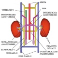
- Paper - The origin of the renal artery in mammals and its anomalies
- Paper - The Terminal Part of the Wolffian Duct
- Paper - The veins of the Wolffian body in the pig
- Template:Polycystic kidney disease
- Template:Pronephros
R
- Template:Ref-Abbott1930
- Template:Ref-Altschule1930
- Template:Ref-Angle1918
- Template:Ref-Bell1935
- Template:Ref-Boyden1931
- Template:Ref-Boyden1932
- Template:Ref-Braasch1912
- Template:Ref-Brambell1927b
- Template:Ref-Bremer1915
- Template:Ref-Bremer1916
- Template:Ref-Bremer1918
- Template:Ref-Eisendrath1912
- Template:Ref-Eisendrath1917
- Template:Ref-Eisendrath1925
- Template:Ref-Gage1905
- Template:Ref-Gladstone1915a
- Template:Ref-Gladstone1924b
- Template:Ref-Gruenwald1937
- Template:Ref-Gruenwald1939
- Template:Ref-Gruenwald1943
- Template:Ref-Hain1936
- Template:Ref-Hain1937
- Template:Ref-Hamann1902
- Template:Ref-Hill1905
- Template:Ref-HinmanGibsonKutzmann1924
- Template:Ref-Howland1916
- Template:Ref-Huber1905
- Template:Ref-JacobYusufJacob2012
- Template:Ref-LewisPapez1915
- Template:Ref-MacCallum1902
- Template:Ref-Mall1901a
- Template:Ref-Minot1898
- Template:Ref-Monie1946
- Template:Ref-MortonJones1936
- Template:Ref-Nagel1889
- Template:Ref-O'Connor1940a
- Template:Ref-O'Connor1940b
- Template:Ref-Oconnor1938
- Template:Ref-OConnor1939
- Template:Ref-Oppenheimer1934
- Template:Ref-PapinEisendrath1927
- Template:Ref-Patten1952b
- Template:Ref-Piersol1927
- Template:Ref-Pohlman1905a
- Template:Ref-Pohlman1905b
- Template:Ref-Pohlman1919
- Template:Ref-Randall1937
- Template:Ref-Rienhoff1922
- Template:Ref-RuscheBacon1939
- Template:Ref-Shikinami1926
- Template:Ref-Siddiqi1937
- Template:Ref-SmithStrasberg1946
- Template:Ref-Spicer1909
- Template:Ref-Stoerk1904
- Template:Ref-Torrey1954
- Template:Ref-Vermooten1951
- Template:Ref-Wakeley1915
- Template:Ref-West1925
- REI - Reproductive Medicine Seminar 2018
- Template:Renal
- Template:Renal abnormalities
- Renal Blood Supply Movie
- Template:Renal drug clearance table
- Template:Renal Histology
- Template:Renal histology
- Template:Renal Links
- Renal System - Abnormalities
- Renal System - Carnegie Stage 13
- Renal System - Carnegie Stage 22
- Renal System - Fetal
- Renal System - Molecular
- Renal System Development
- Renal System Histology
- Template:Renal terms
- Template:Renal Vascular Anomalies
- Royal Hospital for Women - Reproductive Medicine Seminar 2018
U
Media in category 'Renal'
The following 200 files are in this category, out of 283 total.
(previous page) (next page)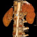 Accessory renal artery.jpg 800 × 798; 103 KB
 Adult bladder.jpg 520 × 273; 34 KB
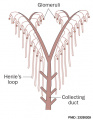 Adult nephron distribution.jpg 502 × 649; 56 KB
 Adult renal venous cartoon.jpg 600 × 600; 62 KB
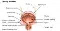
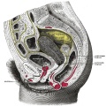
 Adult urinary bladder.jpg 1,164 × 1,000; 148 KB
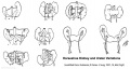
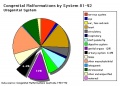 Australian abnormalities 81-92 urogenital.jpg 600 × 429; 54 KB
 Bailey305.jpg 931 × 440; 82 KB
 Bailey306.jpg 624 × 419; 35 KB
 Bailey307.jpg 832 × 833; 128 KB
 Bailey308.jpg 791 × 848; 88 KB
 Bailey309.jpg 594 × 592; 58 KB
 Bailey311.jpg 758 × 578; 50 KB
 Bailey312.jpg 823 × 839; 82 KB
 Bailey313.jpg 543 × 649; 79 KB
 Bailey314.jpg 455 × 461; 25 KB
 Bailey315.jpg 689 × 672; 121 KB
 Bailey316.jpg 671 × 325; 58 KB
 Bailey317-319.jpg 663 × 1,080; 99 KB
 Bailey320.jpg 869 × 582; 75 KB
 Bailey321.jpg 596 × 671; 77 KB
 Bailey322.jpg 766 × 476; 62 KB
 Bailey323.jpg 653 × 521; 50 KB
 Bailey324 325.jpg 706 × 888; 114 KB
 Bailey324.jpg 913 × 558; 78 KB
 Bailey325.jpg 876 × 636; 101 KB
 Bailey326.jpg 502 × 512; 45 KB
 Bailey327.jpg 872 × 567; 89 KB
 Bailey333.jpg 830 × 445; 74 KB
 Bailey340.jpg 814 × 657; 62 KB
 Bladder exstrophy - female.jpg 1,200 × 900; 112 KB
 Bladder exstrophy - male.jpg 637 × 480; 31 KB
 Bladder Exstrophy.jpg 600 × 383; 37 KB
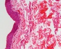
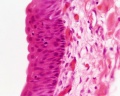
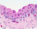
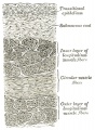
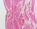 Bladder histology 001.jpg 1,280 × 1,024; 522 KB
 Bladder histology 002.jpg 1,280 × 1,024; 295 KB
 Bladder histology 003.jpg 1,280 × 1,024; 229 KB
 Bladder histology 004.jpg 1,280 × 1,024; 212 KB
 Bladder histology 01.jpg 480 × 600; 29 KB
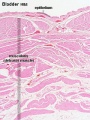 Bladder histology.jpg 300 × 400; 56 KB
 Boyden1931 fig02.jpg 773 × 860; 75 KB
 Boyden1931 fig03.jpg 541 × 878; 56 KB
 Boyden1931 fig04.jpg 1,280 × 652; 130 KB
 Boyden1931 fig05.jpg 1,000 × 646; 97 KB
 Caudal duplication syndrome.jpg 700 × 599; 47 KB
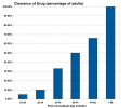 Drug-clearance-rates.png 652 × 542; 14 KB
 Eisendrath1925 fig01.jpg 1,000 × 882; 52 KB
 Eisendrath1925 fig02a.jpg 1,000 × 1,184; 105 KB
 Eisendrath1925 fig04.jpg 1,000 × 983; 85 KB
 Eisendrath1925 fig16.jpg 1,000 × 1,287; 76 KB
 Embryo renal venous cartoon.jpg 600 × 600; 68 KB
 Female genital and ureter abnormality 01.jpg 766 × 732; 86 KB
 Female genital and ureter abnormality 02.jpg 766 × 733; 78 KB
 Female genital and ureter abnormality 03.jpg 766 × 762; 79 KB
 Fetal 10wk urogenital 1.jpg 800 × 600; 109 KB
 Fetal 10wk urogenital 2.jpg 800 × 600; 110 KB
 Fetal 10wk urogenital 3.jpg 800 × 600; 107 KB
 Fetal 10wk urogenital 4.jpg 800 × 600; 105 KB
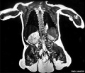
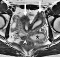
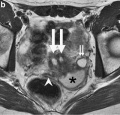
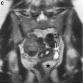
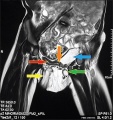
 Fetal kidney MRI 01.jpg 797 × 880; 68 KB
 Fetal kidney MRI 02.jpg 797 × 880; 64 KB
 Fetal kidney.jpg 689 × 623; 43 KB
 Fetal nephron development 01.jpg 1,338 × 689; 119 KB
 Girgis09.jpg 342 × 1,033; 54 KB
 Glomerular podocyte cartoon 02.jpg 1,000 × 280; 51 KB
 Glomerular podocyte cartoon.jpg 800 × 199; 19 KB
 Gray0585.jpg 752 × 800; 188 KB
 Gray0618.jpg 800 × 593; 108 KB
 Gray1111.jpg 523 × 600; 66 KB
 Gray1115.jpg 600 × 474; 73 KB
 Gray1116.jpg 600 × 433; 88 KB
 Gray1117.jpg 581 × 510; 75 KB
 Gray1118.jpg 600 × 403; 45 KB
 Gray1120.jpg 683 × 900; 297 KB
 Gray1121.jpg 600 × 669; 154 KB
 Gray1122.jpg 520 × 404; 57 KB
 Gray1123.jpg 564 × 450; 66 KB
 Gray1124.jpg 714 × 500; 138 KB
 Gray1125.jpg 500 × 584; 69 KB
 Gray1126.png 460 × 500; 34 KB
 Gray1127.jpg 417 × 700; 108 KB
 Gray1128.jpg 700 × 698; 114 KB
 Gray1129.jpg 396 × 500; 55 KB
 Gray1130.jpg 301 × 400; 25 KB
 Gray1131.jpg 90 × 300; 11 KB
 Gray1132.jpg 625 × 500; 86 KB
 Gray1133.jpg 500 × 331; 67 KB
 Gray1137.jpg 600 × 502; 64 KB
 Gray1138.jpg 656 × 472; 57 KB
 Gray1139.jpg 600 × 524; 73 KB
 Gray1140.jpg 600 × 659; 100 KB
 Gray1141.jpg 439 × 600; 91 KB
 Gray1166.jpg 750 × 750; 194 KB
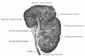
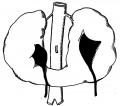
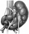
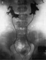
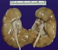 Horseshoe kidney 01.jpg 701 × 600; 68 KB
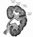
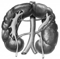
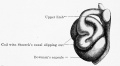
 Horseshoe kidney.jpg 776 × 416; 39 KB
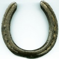 Horseshoe.jpg 400 × 400; 32 KB
 Human bilateral renal agenesis-hypoplasia-dysplasia.png 1,613 × 906; 2.75 MB
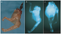
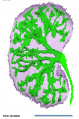
 Human embryonic renal branching 1.jpg 1,280 × 779; 236 KB
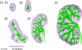
 Human embryonic renal branching stage 22.jpg 500 × 756; 150 KB
 Human fetal kidney histology 01.jpg 1,280 × 1,024; 481 KB
 Human fetal kidney histology 02.jpg 1,280 × 1,024; 322 KB
 Human fetal kidney histology 03.jpg 1,280 × 1,024; 333 KB
 Human fetal kidney histology 04.jpg 1,280 × 1,024; 307 KB
 Human- fetal week 10 lower body A.jpg 600 × 450; 96 KB
 Human- fetal week 10 lower body B.jpg 600 × 450; 93 KB
 Human- fetal week 10 lower body C.jpg 600 × 450; 94 KB
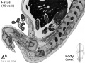
 Human- fetal week 10 urogenital A.jpg 600 × 450; 109 KB
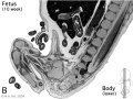
 Human- fetal week 10 urogenital B.jpg 600 × 450; 109 KB
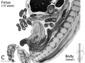
 Human- fetal week 10 urogenital C.jpg 600 × 450; 105 KB
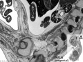
 Human- fetal week 10 urogenital D.jpg 600 × 450; 101 KB
 Hydrocolpos.jpg 375 × 361; 23 KB
 Hydronephrosis.jpg 600 × 345; 64 KB
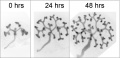
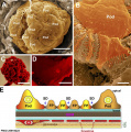
 In vitro kidney development.png 600 × 412; 1.08 MB
 Keibel Mall 2 416.jpg 1,000 × 738; 77 KB
 Keibel Mall 2 520.jpg 1,280 × 998; 167 KB
 Keibel Mall 2 523.jpg 1,280 × 1,234; 208 KB
 Keibel Mall 2 524.jpg 1,280 × 1,072; 208 KB
 Keibel Mall 2 525.jpg 1,280 × 896; 160 KB
 Keibel Mall 2 526.jpg 1,280 × 1,391; 186 KB
 Keibel Mall 2 528.jpg 1,280 × 1,014; 214 KB
 Keibel Mall 2 529.jpg 1,280 × 1,223; 293 KB
 Keibel Mall 2 548.jpg 672 × 483; 53 KB
 Keibel Mall 2 552.jpg 1,280 × 948; 257 KB
 Keibel Mall 2 562.jpg 1,280 × 1,450; 510 KB
 Keibel Mall 2 571.jpg 1,280 × 820; 172 KB
 Keibel Mall 2 572.jpg 1,280 × 969; 219 KB
 Keibel Mall 2 573.jpg 1,280 × 886; 165 KB
 Keibel Mall 2 574.jpg 1,280 × 1,902; 327 KB
 Keibel Mall 2 575.jpg 1,000 × 1,288; 120 KB
 Keibel Mall 2 576.jpg 1,280 × 767; 154 KB
 Keibel Mall 2 578.jpg 1,280 × 836; 115 KB
 Keibel Mall 2 579.jpg 800 × 299; 25 KB
 Keibel Mall 2 583.jpg 1,280 × 1,044; 259 KB
 Keibel Mall 2 584.jpg 464 × 800; 68 KB
 Keibel Mall 2 585.jpg 1,280 × 1,144; 277 KB
 Keibel Mall 2 590.jpg 853 × 702; 66 KB
 Keibel Mall 2 591.jpg 1,004 × 788; 90 KB
 Keibel Mall 2 592.jpg 1,000 × 554; 75 KB
 Keibel Mall 2 596.jpg 1,280 × 1,074; 120 KB
 Keibel Mall 2 598.jpg 1,280 × 854; 146 KB
 Keibel Mall 2 652.jpg 1,200 × 1,036; 130 KB
 Keibel Mall 2 658a.jpg 1,127 × 1,200; 103 KB
 Keibel Mall 2 658b.jpg 895 × 1,200; 98 KB
 Keibel Mall 2 658c.jpg 1,000 × 1,019; 90 KB
 Keibel Mall 2 Felix-plate01.jpg 1,280 × 632; 320 KB
 Keith1902 fig079.jpg 742 × 800; 78 KB
 Keith1902 fig080.jpg 651 × 700; 79 KB
 Keith1902 fig081.jpg 818 × 800; 113 KB
 Keith1902 fig082.jpg 924 × 800; 98 KB
 Keith1902 fig083.jpg 782 × 700; 79 KB
 Keith1902 fig084.jpg 732 × 800; 88 KB
 Keith1902 fig085.jpg 800 × 590; 78 KB
 Keith1902 fig086.jpg 842 × 700; 84 KB
 Keith1902 fig087.jpg 800 × 613; 90 KB
 Keith1902 fig088.jpg 788 × 1,000; 92 KB
 Keith1902 fig089.jpg 964 × 800; 92 KB
 Keith1902 fig090.jpg 700 × 450; 58 KB
 Keith1902 fig091.jpg 700 × 423; 48 KB
 Keith1902 fig093.jpg 700 × 574; 59 KB
 Keith1902 fig094.jpg 800 × 646; 102 KB
 Keith1902 fig095.jpg 660 × 1,000; 125 KB
 Keith1902 fig096.jpg 700 × 535; 69 KB
 Keith1902 fig097.jpg 680 × 592; 80 KB
 Keith1902 fig098.jpg 560 × 460; 40 KB
 Keith1902 fig099.jpg 784 × 800; 105 KB
 Keith1902 fig100.jpg 1,000 × 569; 81 KB
 Keith1902 fig101.jpg 670 × 545; 69 KB
 Keith1902 fig102.jpg 800 × 605; 68 KB
 Keith1902 fig103.jpg 1,000 × 723; 139 KB
 Keith1902 fig104.jpg 800 × 601; 77 KB
 Keith1902 fig105.jpg 632 × 700; 66 KB
 Keith1902 fig108.jpg 780 × 585; 72 KB
 Keith1902 fig109.jpg 859 × 800; 104 KB
 Keith1902 fig110.jpg 800 × 638; 102 KB
 Keith1902 fig111.jpg 1,000 × 620; 145 KB
 Keith1902 fig112.jpg 600 × 541; 77 KB
 Kollmann426.jpg 500 × 620; 46 KB
 Kollmann554.jpg 625 × 568; 54 KB
 Male histology 003.jpg 1,280 × 1,024; 703 KB
 Male histology 004.jpg 1,280 × 1,024; 540 KB
 Male vas deferens and bladder week6to10.jpg 800 × 918; 86 KB
 Mesonephric duct position week 6-11.jpg 697 × 800; 72 KB
 Mouse Kidney Development Cartoon.jpg 506 × 658; 120 KB
 Mouse nephron stages 01.jpg 601 × 916; 217 KB
 Mouse renal podocyte EM01.jpg 1,000 × 1,338; 366 KB
 Mouse renal podocyte EM02.jpg 1,000 × 666; 155 KB
 Mouse-kidney in vitro.jpg 955 × 461; 57 KB
 Mouse-kidney model GDNF-FGF10.jpg 405 × 928; 59 KB
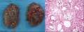 Multicystic kidney and histology.jpg 1,000 × 384; 108 KB
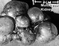 Multicystic kidney.jpg 600 × 468; 56 KB
 Multiple renal arteries 01.jpg 496 × 496; 40 KB
 Neonatal duplicated bladder MRI 01.jpg 751 × 800; 116 KB
 Nephron development 01.jpg 1,232 × 960; 240 KB
 Nephron EM01.jpg 1,909 × 1,280; 219 KB
 Nephron EM02.jpg 1,271 × 1,280; 210 KB
 Nephron EM11.jpg 2,983 × 2,000; 398 KB
 Nephron histology 01.jpg 400 × 500; 79 KB
 Nephron histology 02.jpg 400 × 500; 77 KB
 Nephron histology 03.jpg 375 × 500; 97 KB
 Nephron histology 04.jpg 375 × 500; 54 KB
 Nephron histology.jpg 400 × 500; 70 KB
 Nephron physiology.jpg 381 × 262; 49 KB
 Nephrons-cortical and juxtamedullary.jpg 507 × 600; 39 KB


