Category:Pfannenstiel III

This Embryology category shows pages and media related to the historic human embryo Pfannenstiel III, named after Professor Pfannenstiel of Griefswald.
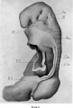
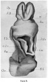
Corresponding to Carnegie stage 11 development in week 4, 23 - 26 days, GA week 6-7. This embryo stage has a crown rump length (CRL) of 2.5 - 4.5 mm and somite number 13 - 20 pairs.
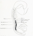
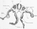
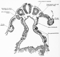
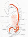
Pfannenstiel III (14 somites) Carnegie stage 11
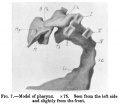
Described by Keibel F., and Elze, C. 1908. Normentafeln zur Entwicklungsgeschichte der Wirbeltiere. (Normal Plates for Evolution of Vertebrates) 8. Heft Normentafeln zur Entwicklungsgeschichte des Menschen. (Vol. 8. Normal Plates of the Development of the Human Embryo) Fisher, Jena., Germany. Shown as Embryo No.6 (Plate 1 fig. Vr. and Plate 2 fig. Vv.).
Described also by Low, A. in 1908.[1]
- Plastic models available of - external form, central nervous system, digestive system, heart and large vessels.
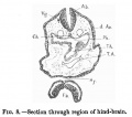
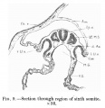
- two pharyngeal arches, clearly marked.
- neural lips are closed rostrally as far as the collicular region.
Described also by Keibel F. in the textbook Keibel F. The Interdependence of the Various Developmental Processes in Keibel F. and Mall FP. Manual of Human Embryology II. (1912) J. B. Lippincott Company, Philadelphia. Chapter XX.
The Interdependence of the Various Developmental Processes
| Human Embryo Pfannenstiel III | ||||||||||||||||||||||
|---|---|---|---|---|---|---|---|---|---|---|---|---|---|---|---|---|---|---|---|---|---|---|
| Collection of Prof. Pfannenstiel, Giessen. Carnegie stage 11 | ||||||||||||||||||||||
| Desig. | Size | Age | Body form | Primitive streak | Primitive segments | Chorda | Nervous system | Eye | Ear | Nose | Hypophysis | Mouth | Digestive trac, liver and pancreas | Branchial pouches, threoid thymus, trachea and lungs | Urogenital system | Heart and vessels | Integument | Skeleton | Extremities | Amnion | Allantois | Remarks |
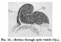
| 6 Pfannenstiel III Fig. Vr and Vv. Textfig. 6a to 6w. | Gr. L. about 2.6 mm. | Embryo bent over the ventral surface and slightly bent spirally. | Tail bud, on its ventral side doubtful remains of primitive streak. | 13-14 pairs of primitive segments. | Chorda emerging from entoderm, Cranially is still entoderm, caudally it is probably primarily independent of the entoderm and in this region it is no longer included in the entoderm. | In brain region medullary canal is open to caudal to the region of the optic vesicle, similarly the caudal end. Anlagen of neuromeres already present. | Primary optic vesicles. They are close to the ectoderm; in their region the medullary canal is wide open. | Anlage of auditory vesicle recognizable as a thickened and at first but little depressed plate of ectoderm. | Hypophysis just indicated. | Primary pharyngeal membrane still closed. Oral sinus. | Wide hepatic bay just cranial to the intestinal umbilicus. No trace yet of hepatic trabeculae. | The two first branchial pouches reach the ectoderm, the 3rd is formed. | Quite rudimentary "pronephric anlage" in 8th, 9th and 10 pairs of primitive segments. No trace yet of a Wolffian duct. Segmental vesicles in the 11th, 12th and 13th pairs of primitive segments. | Heart S-shaped. Posterior mesocardium through a few sections. Aorta paired throughout. | Allantoic duct | Extirpation of uterus on account of carcinoma. | ||||||
| Remarks - Fixation formalin Muller's fluid. Stain Paracarmine. Sections 10 μ. Recent mitoses. Septum transverse. No ventral connection between pericardial and peritoneal cavities. | ||||||||||||||||||||||
| Reference: Keibel F. and Elze C. Normal Plates of the Development of the Human Embryo (Homo sapiens). (1908) Vol. 8 in series by Keibel F. Normal plates of the development of vertebrates (Normentafeln zur Entwicklungsgeschichte der Wirbelthiere) Fisher, Jena., Germany. Cited in: 1912 Human Embryology and Low A. Description of a human embryo of 13-14 mesodermic somites. (1908) J Anat Physiol. 42(3): 237-51. PMID 17232769 | PMC1289161 | ||||||||||||||||||||||
| 1908 Embryo Tables: Klb (stage 10) | Pfannenstiel III (stage 11) | Meyer 300 (stage 12) | Strahl 4mm (stage 13) | Hertwig G31 (stage 14) | ||||||||||||||||||||||
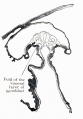
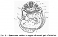
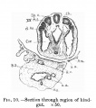
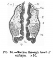
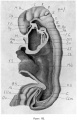
Fig. 600 Reconstruction of the caudal end of the body of Embryo Pfannenstiel III 2.6 mm greatest length and with 13-14 pairs of primitive segments. X 50.
References
- ↑ Low A. Description of a human embryo of 13-14 mesodermic somites. (1908) J Anat Physiol. 42(3): 237-51. PMID 17232769 | PMC1289161
| Week: | 1 | 2 | 3 | 4 | 5 | 6 | 7 | 8 |
| Carnegie stage: | 1 2 3 4 | 5 6 | 7 8 9 | 10 11 12 13 | 14 15 | 16 17 | 18 19 | 20 21 22 23 |
- Carnegie Stages: 1 | 2 | 3 | 4 | 5 | 6 | 7 | 8 | 9 | 10 | 11 | 12 | 13 | 14 | 15 | 16 | 17 | 18 | 19 | 20 | 21 | 22 | 23 | About Stages | Timeline
| Carnegie Collection - Stage 11 | ||||||||||
|---|---|---|---|---|---|---|---|---|---|---|
| Serial No. | Pairs of somites | Size (mm) | Grade | Fixative | Embedding Medium | Plane | Thinness (µm) | Stain | Year | Notes |
| 12 | 14 | E, 2.1 Ch, 13 | Poor | P | Transverse | 10 | Al. carm. | 1893 | ||
| 164 | 18 | E, 3.5 Ch, 14 | Good | Formalin | P | Transverse | 20 | Al. carm. | 1913 | |
| 318 | 13/14 | E, 2.5 Ch, 16 | Good | P | Transverse | 25 | Al. carm. | 1905 | ||
| 470 | 17 | E, 4.3 Ch, 16 | Good | Formalin | P | Transverse | 10 | Al. carm. . | 1910 | |
| 779 | 14 | E, 2.75 | Good | C | Transverse | 15 | Al. coch. | 1913 | Dysraphism. Noted by Dekaban (1964)[1] | |
| 1182b | E, 3 Ch, 15x12x5 | Good | Formalin | ? | Transverse | 20 | Al. carm. | 1915 | ||
| 2053 | 20 | E, 3.1 Ch, 12 | Exc. | Formalin | P | Transverse | 10 | Al. coch. | 1918 | Most advanced in group. Ag added to slide 2 Monographs by Davis (1923)[2] and Congdon (1922)[3] |
| 4315 | 17 | E, 4.7 Ch, 23x10.4X11 | Excellent | ? | C-P | Transverse | 10 | I.H. & E. | 1923 | Univ. Chicago No. 951. Wen (1928)[4] |
| 4529 | 14 | E, 2.4 Ch, 21 | Excellent | Formalin | P | Transverse | 10 | Al. coch, or. G. | 1924 | Heuser (1930)[5] |
| 4783 | 13 | E, 2.3 | Fair | ? | ? | Transverse | 5 | I.H. | 1924 | Wallin (1913)[6] |
| 4877 | 13 | E, 2 Ch, 15 | Good | Formalin | P | Transverse | 15 | Al. coch. | 1925 | |
| 5072 | 17 | E, 3 | Good | Formalin | P | Transverse | 10 | (Stain - Haematoxylin Eosin) | 1925 | Tubal Type specimen. Atwell (1930)[7] |
| 6050 | 19/21 | E.,3 Ch, 10 | Good | Formalin | C-P | Coronal | 10 | Al. coch. | 1930 | Advanced |
| 6344 | 13 | E, 2.5 Ch, 17 | Excellent | Formalin | C-P | Transverse | 6 | Al. coch. | 1931 | Least advanced in group |
| 6784 | 17 | E, 5 Ch, 16 | Excellent | Formalin | C-P | Transverse | 6 | I.H, or. G. | 1933 | |
| 7358 | 16 | E, ? Ch, 15 | Poor | Alc, formol | p | Oblique | 25 | (Stain - Haematoxylin Eosin) | 1936 | |
| 7611 | 16 | E., 2.4 Ch., 12 | Excellent | Bouin | C-P | Transverse | 8 | (Stain - Haematoxylin Eosin) | 1938 | |
| 7665 | 19 | E., 4.36 | Excellent | ? | C-P | Transverse | 6 | 1939 | Univ. Chicago No. H 1516 | |
| 7702 | 17 | E, 3.7 Ch., 14 | Good | Formalin | C-P | Transverse | 10 | Al. coch. | 1940 | Returned to B M Patten |
| 7851 | 13 | E., 4.3 Ch, 18 | Excellent | Formalin | C-P | Transverse | 8 | (Stain - Haematoxylin Eosin) | 1940 | Slightly injured |
| 8005 | 16/17 | E, 3 | Excellent | Bouin | C-P | Transverse | 8 | (Stain - Haematoxylin Eosin) | 1942 | Tubal |
| 8116 | 17 | E, 14 Ch.. 17 | Good | Formalin | p | Sagittal | 8 | Azan | 1953 | |
| 8962 | 15 | E, 1.55 | Good | ? | * | Sagittal | ? | ? | 1952 | Tubal Univ. Chicago No. H 810 |
Abbreviations
| ||||||||||
References
| ||||||||||
Pages in category 'Pfannenstiel III'
The following 7 pages are in this category, out of 7 total.
Media in category 'Pfannenstiel III'
The following 24 files are in this category, out of 24 total.
 Keibel Mall 2 526.jpg 1,280 × 1,391; 186 KB
 Keibel Mall 2 528.jpg 1,280 × 1,014; 214 KB
 Keibel Mall 2 529.jpg 1,280 × 1,223; 293 KB
 Keibel Mall 2 541.jpg 1,280 × 1,683; 313 KB
 Keibel Mall 2 545.jpg 761 × 1,093; 104 KB
 Keibel Mall 2 600.jpg 1,000 × 692; 77 KB
 Keibel Mall 2 609.jpg 1,200 × 753; 130 KB
 Low 01.jpg 502 × 535; 60 KB
 Low 02.jpg 900 × 328; 58 KB
 Low 04.jpg 535 × 541; 40 KB
 Low 05.jpg 529 × 552; 45 KB
 Low 06.jpg 751 × 484; 59 KB
 Low 07.jpg 711 × 619; 56 KB
 Low 08.jpg 566 × 499; 48 KB
 Low 09.jpg 589 × 609; 50 KB
 Low 10.jpg 519 × 586; 61 KB
 Low 11.jpg 414 × 451; 49 KB
 Low 12.jpg 450 × 419; 43 KB
 Low 13.jpg 637 × 443; 52 KB
 Low 14.jpg 508 × 554; 60 KB
 Low 15.jpg 565 × 570; 73 KB
 Low plate 01.jpg 661 × 981; 137 KB
 Low plate 02.jpg 581 × 980; 118 KB
 Low plate 03.jpg 707 × 1,099; 166 KB


