Category:Middle Ear
From Embryology
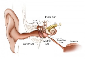
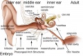
This Embryology category shows pages and media related to the middle ear development.
- Links: Middle Ear Development
Pages in category 'Middle Ear'
The following 35 pages are in this category, out of 35 total.
P
- Paper - Development of the aquaeductus cochleae and its contained periotic duct and cochlear vein in human embryos
- Paper - Development of the incus of the human ear - illustrated in atlas series
- Paper - Development of the malleus of the human ear - Illustrated in atlas series
- Paper - Development of the otic capsule of the human ear - illustrated in atlas series
- Paper - Development of the stapes of the human ear - illustrated in atlas series
- Paper - Major features in the developmental history of the human stapes (1940)
- Paper - Postnatal growth and adult structure of the otic (endolymphatic) sac
- Paper - Stapes, fissula ante fenestram and associated structures in man 1
- Paper - Stapes, fissula ante fenestram and associated structures in man 2
- Paper - Stapes, fissula ante fenestram and associated structures in man 5
- Paper - The development of the auditory ossicles and associated structures in man
- Paper - The development of the auditory ossicles, the otic capsule and the extracapsular tissues
- Paper - The development of the ear-bones in the mouse
- Paper - The development of the otic capsule in the region of surgical fenestration 1
- Paper - The development of the otic capsule in the region of surgical fenestration 2
- Paper - The early embryology of the auditory ossicles in man
- Paper - The early formations of the middle ear and eustachian tube - a criticism
R
Media in category 'Middle Ear'
The following 38 files are in this category, out of 38 total.
 Adult hearing embryonic origins.jpg 1,000 × 675; 80 KB
 Anson1948 fig01.jpg 1,280 × 1,635; 310 KB
 Anson1948 fig02.jpg 1,280 × 1,232; 241 KB
 Anson1948 fig03.jpg 1,280 × 1,815; 358 KB
 Anson1948 fig04.jpg 1,280 × 1,541; 247 KB
 Anson1948 fig05.jpg 1,280 × 1,797; 362 KB
 Anson1948 fig06.jpg 1,028 × 1,488; 224 KB
 Anson1948 fig07.jpg 1,280 × 1,801; 219 KB
 Anson1948 fig08.jpg 1,280 × 1,764; 207 KB
 Anson1948 fig09.jpg 1,280 × 1,686; 235 KB
 Anson1948 fig10.jpg 1,191 × 1,236; 204 KB
 Anson1948 fig11.jpg 1,220 × 981; 230 KB
 Anson1948 fig12.jpg 1,280 × 1,490; 289 KB
 Anson1948 fig13.jpg 1,239 × 841; 203 KB
 Anson1948 fig16.jpg 1,280 × 817; 133 KB
 AnsonKarabinMartin1939 fig01-06.jpg 1,280 × 1,650; 324 KB
 AnsonKarabinMartin1939 fig07-12.jpg 1,280 × 1,880; 444 KB
 AnsonKarabinMartin1939 fig13-15.jpg 1,280 × 1,519; 171 KB
 AnsonKarabinMartin1939 fig60-64.jpg 1,280 × 1,496; 130 KB
 BeatonAnson1940 fig01-2.jpg 1,652 × 2,147; 652 KB
 Eustacian tube angle.jpg 609 × 458; 48 KB
 Frazer1922 fig01.jpg 627 × 900; 102 KB
 Frazer1922 fig02.jpg 1,000 × 853; 182 KB
 Frazer1922 fig03.jpg 1,000 × 697; 165 KB
 Frazer1922 fig04.jpg 1,000 × 927; 136 KB
 Frazer1922 fig05.jpg 1,000 × 783; 94 KB
 Frazer1922 fig06.jpg 1,000 × 728; 116 KB
 Gray0909.jpg 600 × 437; 56 KB
 Gray0910.jpg 400 × 548; 78 KB
 Gray0916.jpg 600 × 367; 28 KB
 Gray0917.jpg 600 × 379; 44 KB
 Gray0918.jpg 500 × 283; 17 KB
 Gray0919.jpg 500 × 731; 93 KB
 HansonAnson1962 fig01.jpg 1,280 × 643; 218 KB
 HansonAnson1962 fig02.jpg 1,280 × 601; 233 KB
 HansonAnson1962 fig04.jpg 1,280 × 598; 201 KB
 HansonAnson1962 fig05.jpg 1,280 × 604; 225 KB
 Meckels cartilage - middle ear from the jaw.jpg 1,161 × 1,280; 259 KB


