Category:Integumentary
From Embryology
This Embryology category shows pages and media related to Integumentary System Development. This includes related topics and undergraduate classes as well as pages and sub-categories describing specific components.
- Ectoderm - epidermis, glands, hair follicles
- Neural Crest - melanocytes
- Mesoderm - dermis, hypodermis, hair follicles
| Integumentary Terms | ||
|---|---|---|
Integumentary Development
| ||
|
Subcategories
This category has the following 7 subcategories, out of 7 total.
Pages in category 'Integumentary'
The following 91 pages are in this category, out of 91 total.
A
B
H
I
- Template:ICD Integumentary (Q80-Q89)
- Template:Integumentary
- Template:Integumentary abnormalities
- Template:Integumentary gland
- Template:Integumentary Links
- Integumentary System - Abnormalities
- Integumentary System - Eyelid Development
- Integumentary System - Gland Development
- Integumentary System - Hair Development
- Integumentary System - Histology
- Integumentary System - Mammary Gland Development
- Integumentary System - Nail Development
- Integumentary System - Tooth Development
- Integumentary System Development
- Integumentary System Development - Vernix Caseosa
- Template:Integumentary terms
M
P
- Template:Pacinian corpuscle
- Paper - Development of the mammary gland
- Paper - Development of the mammary gland - Arris and Gale Lecture
- Paper - On the role of the developing epidermis in forming sheaths and lumina to organs
- Paper - Sensory nerves in the skin of human fetuses of 8 to 14 weeks of menstrual age correlated with functional capability (1941)
- Paper - Studies on the mammary gland 2 (1917)
- Paper - The development and evolution of the "papillary" ridges and patterns on the volar surface of the hand (1906)
- Paper - The development of the eyelids 1
- Paper - The development of the subcutaneous vascular plexus in the head of the human embryo (1923)
- Paper - The relations of endogenous and exogenous factors in bone and tooth development (1937)
- Paper - The weight of the skin and tela subcutanea of the human fetus
- Paper The development of the subcutaneous vascular plexus in the head of the human embryo (1923)
- Template:Prepuce
R
S
Media in category 'Integumentary'
The following 172 files are in this category, out of 172 total.
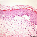
 Adult epidermis histology 01.jpg 600 × 750; 83 KB
 Adult epidermis histology 02.jpg 600 × 750; 123 KB
 Adult skin histology 01.jpg 600 × 750; 73 KB
 Adult skin histology 02.jpg 600 × 750; 88 KB
 Adult skin histology 03.jpg 600 × 750; 108 KB
 Apocrine secretion animation.gif 60 × 80; 4 KB
 Arey1924 fig229.jpg 1,000 × 625; 150 KB
 Arey1924 fig230.jpg 1,000 × 707; 117 KB
 Arey1924 fig231.jpg 1,200 × 748; 188 KB
 Arey1924 fig233.jpg 1,200 × 597; 156 KB
 Arey1924 fig234.jpg 1,200 × 908; 214 KB
 Arey1924 fig235.jpg 1,200 × 523; 157 KB
 Bailey110.jpg 481 × 643; 110 KB
 Bailey353.jpg 759 × 458; 60 KB
 Bailey354.jpg 866 × 484; 61 KB
 Bailey355.jpg 632 × 890; 109 KB
 Bailey356.jpg 767 × 533; 103 KB
 Bailey357.jpg 949 × 395; 88 KB
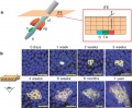
 Chicken skin timeline 01.jpg 1,547 × 1,709; 218 KB
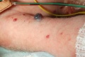
 Cutis aplasia.jpg 500 × 374; 24 KB
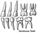 Deciduous teeth.jpg 250 × 220; 13 KB
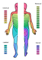 Dermatomes.png 424 × 600; 82 KB
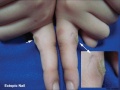 Ectopic nail.jpg 800 × 603; 70 KB
 Elaine Fuchs.jpg 471 × 393; 135 KB
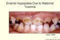 Enamel Hypoplasia Due to Maternal Toxemia.jpg 700 × 466; 38 KB
 Epidermis cartoon 01.jpg 545 × 375; 96 KB
 Epidermis cartoon 02.jpg 452 × 536; 78 KB
 Epidermis cartoon.jpg 800 × 536; 126 KB
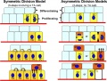 Epidermis-stem cell models.jpg 800 × 605; 111 KB
 Epidermolysis bullosa simplex 01.jpg 498 × 498; 62 KB
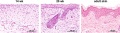 Fetal integumentary histology 01.jpg 800 × 219; 74 KB
 Fetal integumentary histology 02.jpg 600 × 664; 145 KB
 Fingernail cartoon.jpg 400 × 214; 15 KB
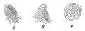
 Fingerprint.jpg 200 × 170; 12 KB
 Gray0892.jpg 500 × 409; 47 KB
 Gray0893.jpg 355 × 700; 93 KB
 Gray0894.jpg 710 × 400; 74 KB
 Gray0895.jpg 700 × 452; 94 KB
 Gray0896.jpg 600 × 638; 68 KB
 Gray0897.jpg 500 × 366; 47 KB
 Gray0940.jpg 618 × 700; 160 KB
 Gray0941.jpg 700 × 524; 106 KB
 Gray0942.jpg 800 × 515; 127 KB
 Gray0943.jpg 800 × 277; 71 KB
 Gray0944.jpg 800 × 681; 178 KB
 Gray0945.jpg 400 × 585; 76 KB
 Gray0946.jpg 500 × 427; 85 KB
 Hair development stages.jpg 1,000 × 590; 159 KB
 Hair development stages.png 600 × 354; 158 KB
 Hair follicle cell development.png 2,012 × 681; 1.44 MB
 Hair follicle development 01.jpg 1,000 × 611; 182 KB
 Hair follicle development 02.jpg 800 × 616; 75 KB
 Hair follicle development.jpg 800 × 663; 191 KB
 Hair histology.jpg 600 × 451; 131 KB
 Holocrine secretion animation.gif 60 × 80; 16 KB
 Human embryo skin 24 week EGA.jpg 596 × 939; 165 KB
 Human embryo skin 8-9 week EGA desmosomes.jpg 800 × 198; 40 KB
 Human embryo skin 8-9 week EGA.jpg 657 × 872; 188 KB
 Human embryo skin 9-11 week EGA.jpg 623 × 804; 176 KB
 Human- Stage 22 integument 01.jpg 1,000 × 750; 205 KB
 Human- Stage 22 integument 02.jpg 800 × 600; 147 KB
 Human- Stage 22 integument 03.jpg 600 × 450; 95 KB
 Human- Stage 22 integument 04.jpg 400 × 300; 48 KB
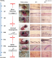
 Inherited dentine disorders.jpg 600 × 903; 98 KB
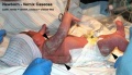
 Integumentary - lip01.jpg 1,280 × 1,024; 253 KB
 Integumentary histology 01.jpg 480 × 600; 70 KB
 Integumentary histology 02.jpg 480 × 600; 58 KB
 Integumentary histology 03.jpg 480 × 600; 85 KB
 Integumentary histology 04.jpg 280 × 700; 65 KB
 Integumentary histology 10.jpg 800 × 1,000; 101 KB
 Integumentary System Fetal Development Timeline.jpg 1,611 × 604; 366 KB
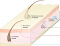
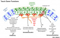 Integumentary touch dome functions.jpg 893 × 556; 97 KB
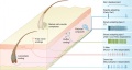
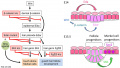 Integumentary touch dome model 01.jpg 1,280 × 724; 144 KB
 Integumentary touch dome model 02.jpg 675 × 724; 73 KB
 Integumentary touch dome model 03.jpg 603 × 724; 73 KB
 Integumentary- hair follicle 01.jpg 479 × 599; 66 KB
 Integumentary- hair follicle 02.jpg 479 × 599; 74 KB
 Integumentary- hair follicle 03.jpg 479 × 599; 48 KB
 Integumentary- sebaceous gland histology 01.jpg 400 × 500; 125 KB
 Integumentary- sebaceous gland histology 02.jpg 400 × 500; 150 KB
 Keibel Mall 196.jpg 448 × 383; 35 KB
 Keibel Mall 197.jpg 500 × 469; 46 KB
 Keibel Mall 198.jpg 500 × 443; 57 KB
 Keibel Mall 199.jpg 800 × 487; 61 KB
 Keibel Mall 200.jpg 781 × 645; 74 KB
 Keibel Mall 201.jpg 434 × 543; 65 KB
 Keibel Mall 202.jpg 481 × 500; 47 KB
 Keibel Mall 203-205.jpg 838 × 1,000; 129 KB
 Keibel Mall 206.jpg 415 × 750; 79 KB
 Keibel Mall 207.jpg 800 × 538; 59 KB
 Keibel Mall 208.jpg 800 × 628; 65 KB
 Keibel Mall 209.jpg 458 × 990; 89 KB
 Keibel Mall 210.jpg 340 × 542; 41 KB
 Keibel Mall 211.jpg 427 × 436; 43 KB
 Keibel Mall 212.jpg 650 × 371; 39 KB
 Keibel Mall 213.jpg 670 × 547; 49 KB
 Keibel Mall 214.jpg 650 × 497; 56 KB
 Keibel Mall 215.jpg 727 × 379; 53 KB
 Keibel Mall 216.jpg 366 × 356; 30 KB
 Keibel Mall 217.jpg 600 × 198; 24 KB
 Keibel Mall 218.jpg 600 × 221; 25 KB
 Keibel Mall 222.jpg 684 × 543; 84 KB
 Keith1902 fig051.jpg 483 × 233; 12 KB
 Keith1902 fig052.jpg 483 × 233; 23 KB
 Keith1902 fig053.jpg 483 × 233; 20 KB
 Keith1902 fig054.jpg 750 × 277; 37 KB
 Keith1902 fig055.jpg 800 × 699; 136 KB
 Keith1902 fig056.jpg 800 × 278; 49 KB
 Keith1902 fig057.jpg 887 × 700; 159 KB
 Keith1902 fig058.jpg 1,172 × 696; 200 KB
 Keith1921 fig490.jpg 1,200 × 656; 214 KB
 Kollmann652.jpg 634 × 374; 31 KB
 Kollmann653.jpg 681 × 400; 56 KB
 Kollmann654.jpg 732 × 584; 72 KB
 Kollmann655-656.jpg 772 × 555; 76 KB
 Kollmann657.jpg 715 × 440; 68 KB
 Kollmann658.jpg 772 × 354; 57 KB
 Kollmann659.jpg 548 × 369; 42 KB
 Kollmann660.jpg 607 × 312; 22 KB
 Kollmann661.jpg 497 × 401; 25 KB
 Kollmann662.jpg 699 × 515; 56 KB
 Kollmann663.jpg 671 × 388; 39 KB
 Kollmann664.jpg 736 × 414; 47 KB
 Kollmann665.jpg 701 × 467; 55 KB
 Kollmann666.jpg 897 × 1,000; 140 KB
 Kollmann667.jpg 646 × 480; 32 KB
 Kollmann668-670.jpg 788 × 566; 35 KB
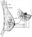 Mammary anatomy.jpg 600 × 702; 58 KB
 Meissner corpuscle 01.jpg 793 × 662; 86 KB
 Meissner corpuscle 02.jpg 800 × 1,000; 67 KB
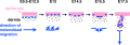 Melanoblast migration.png 600 × 210; 40 KB
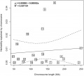 Melanoma chromosome graph.jpg 800 × 727; 76 KB
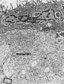
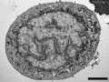 Merkel cell EM 01.jpg 984 × 738; 209 KB
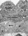
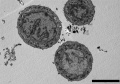 Merkel cell EM 02.jpg 984 × 685; 166 KB
 Merocrine secretion animation.gif 60 × 80; 10 KB
 Mice cloned from adult keratinocytes.jpg 600 × 398; 20 KB
 Mouse mammary development 01.jpg 1,200 × 773; 147 KB
 Mouse melanoblast distribution 01.jpg 697 × 1,000; 192 KB
 Mouse melanoblast distribution 02.jpg 777 × 1,121; 129 KB
 Mouse melanoblast distribution 03.jpg 751 × 1,051; 150 KB
 Mouse melanoblast distribution 04.jpg 761 × 1,128; 177 KB
 Mouse melanoblast distribution 05.jpg 761 × 540; 94 KB
 Mouse melanoblast distribution 06.jpg 761 × 524; 74 KB
 Mouse tooth stem cell.png 600 × 161; 70 KB
 Mouse-mammary-E10-E11.jpg 600 × 406; 40 KB
 Mouse-mammary-E11.5.jpg 600 × 406; 78 KB
 Mouse-mammary-E12.0.jpg 600 × 398; 89 KB
 Mouse-mammary-E13.jpg 600 × 415; 57 KB
 Mouse-mammary-E14.5.jpg 600 × 459; 77 KB
 Nail patella syndrome 01.jpg 543 × 632; 42 KB
 Nail Plate Development -Lewis .jpg 692 × 364; 97 KB
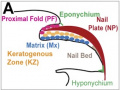 Nail structure cartoon1.jpg 500 × 377; 54 KB
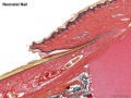 Neonatal nail.jpg 800 × 600; 176 KB
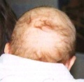 Neonate hair.jpg 320 × 313; 15 KB
 Neuroblastoma.jpg 640 × 426; 31 KB
 Newborn - vernix caseosa.jpg 800 × 463; 73 KB
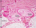 Pacinian corpuscle histology 01.jpg 800 × 640; 227 KB
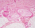 Pacinian corpuscle histology 02.jpg 1,280 × 1,024; 207 KB
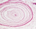 Pacinian corpuscle histology 03.jpg 1,280 × 1,024; 133 KB
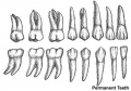 Permanent teeth.jpg 400 × 280; 23 KB
 Permanentteeth.jpg 400 × 280; 23 KB
 Skin structure cartoon.jpg 700 × 720; 52 KB
 Stratified epithelia cartoon.jpg 696 × 1,000; 166 KB
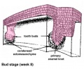
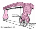 Tooth bell stage.jpg 430 × 352; 35 KB
 Tooth bud stage.jpg 430 × 352; 35 KB
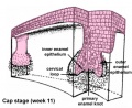 Tooth cap stage.jpg 430 × 352; 38 KB
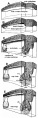 Tooth development stage.jpg 430 × 1,338; 303 KB
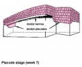 Tooth placode stage.jpg 430 × 352; 22 KB
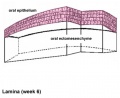 Tooth stage lamina.jpg 430 × 352; 22 KB
 Touch receptors in mammalian skin cartoon 01.jpg 600 × 459; 38 KB
 Touch receptors in mammalian skin cartoon.jpg 800 × 429; 57 KB
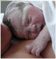 Vernix caseosa 01.jpg 800 × 839; 31 KB


