ANAT2341 Lab 5 - Early Embryo
| Lab 5: Introduction | Trilaminar Embryo | Early Embryo | Late Embryo | Fetal | Postnatal | Abnormalities | Online Assessment |
We have now reached the end of Week 4 and beginning of 5 of development. Start by looking briefly at the overview of the Carnegie stage 13 embryo GIT from one end to the other.
Then work through the listed specific serial sections of the embryo identifying the GIT features. Alternatively step through the serial sections yourself identifying the tract, its associated mesentries, organs and spaces.
Tract Development

|

|
| Early Embryo | Later Embryo |
Carnegie Stage 13 (Week 4-5)

|

| |
| The individual serial slices have also been incorporated into a 3D model of this embryo. |
| Section | Name | Description |
|---|---|---|

|
B1L | Pharynx
The righthand side towards the buccopharyngeal membrane, the lefthand side descending into the embryo body. Central region is the floor of pharynx formed by fusion of 3rd pharyngeal arches = hypopharyngeal eminence (precursor of root of tongue). Rathke's pouch forming the rudimentary adenohypophysis (anterior pituitary). |

|
B3L | Laryngeal tracheal groove - beginning of ventral compression, at 90 degrees to the lateral plane of the pharynx above this point.
Rudimentary thyroid ventral to aortic sac (also seen in B2, ventral to the hypopharyngeal eminence). |

|
B4L | Laryngeal tracheal groove - Caudal pharynx compressed dorsoventrally.
Note that it lies between the aortic sac (ventrally) directly above the heart and the paired vessels of arch artery 6 and the dorsal aortas. The pale staining region behind these blood vessels is where the vertebral column will form. |

|
B5L | Laryngopharynx - now compressed dorsoventrally between the paired arch artery 6 vessels. |

|
B7L | Glottis - initial separation of the oesophagus (dorsal) from the trachea (ventral).
Note that this is occurring at the level of the heart atria. Nasal placodes. Pulmonary arteries. |

|
C1L | Gastrointestinal tract oesophagus (dorsal) is now separate from the respiratory trachea (ventral). |

|
C2L | Oesophagus and trachea both surrounded by dense mesenchyme.
Right nasal pit. |

|
C3L | Oesophagus and trachea both surrounded by dense mesenchyme.
Common cardinal vein in the posterior wall of the intraembryonic coelom. The pleuropericardial folds which contribute later to the formation of the pleura and pericardium. In C4, junction of right common cardinal vein with dorsal wall of sinus venosus. Left nasal pit. |

|
C5L | Smaller oesophagus and expanding trachea, this is also the upper region of the lung buds.
The ventral anchoring of attachment site is at the most cranial extension of the septum transversum. This attachment now divides the intraembryonic coelom around the trachea into two canals, the left and right pleuro (pericardio-peritoneal) canals. Canals are lined by coelomic mesothelium and are continuous with whole intraembryonic coelom (they will be referred to hereafter simply as coelomic canals). The pleuroperitoneal fold on the medial side of the right common cardinal vein will form part of the diaphragm. |

|
C6L | Trachea expanded and beginning to bifurcate to the major bronchial branches for each lung.
Lateral extension of pulmonary mesenchyme is moulded to shape of coelomic canals. Oesophagus lumen obliterated (common site of oesophageal atresia and/or tracheo-oesophageal fistula). Prominent R pleuroperitoneal fold. |

|
C7L | Trachea bifurcated to the major bronchial branches for each lung.
Note dorsal extent of coelomic canals. Oesophagus lumen reappears caudal to bifurcation. Distinct R (smaller on L) pleuroperitoneal fold below the common cardinal vein. |

|
D1L | Oesophagus/stomach junction.
Right lung bud bronchus still visible, left bronchus ends above this section. Note the oesophagus now lies in the midline between the 2 bronchi. Coelomic canals. |

|
D2L | Ovoid stomach with developing space of the lesser sac on R.
Dorsal and ventral attachments of the mesenchyme are now known as dorsal and ventral mesogastria. Coelomic canals. |

|
D3L | Rotation of stomach (seen from above) to right side.
Note change in outline of coelomic canals due to presence of liver. Lesser sac. Note thick mesothelium lining the coelom along left edge of stomach, the primordium of the spleen and greater omentum along greater curvature. Liver embedded in septum transversum (ventral border of septum transversum contributes to diaphragm). |

|
D4L | Rotation of stomach (seen from above) to right side.
Ventral mesogastrium - Stomach is attached ventrally to the liver. (note the position of the ductus venosus) Dorsal mesogastrium - within this structure the spleen will begin to form and later the greater omentum. Peritoneal spaces - identify greater and lesser sac. |

|
D6L | Pyloric region of stomach.
Ventral mesogastrium - Stomach is closely attached ventrally to the liver. Dorsal mesogastrium - within this structure the spleen will begin to form and later the greater omentum. Peritoneal spaces - identify greater and lesser sac. |

|
E4L | Midgut.
Region close to the umbilicus. Note the close associated portal vein and the paired placental (umbilical) veins. |

|
F1L | Midgut.
Looping out of body wall ventrally (cut tangentially). Also note the righthand side hindgut region. |

|
G7L | Caudal pharynx (extending laterally, ventral to dorsal aorta - cf B4). Stomach, mesentery |

|
G6L | Narrow oesophagus. Tracheal bifurcation dorsal to sinus venosus. |


Peritoneal Cavity
- Fusion of the two separate intra-embryonic coelom spaces.
- Intestinal loss of ventral attachment, except at the level of the stomach, dorsal attachment becomes true mesentery.
Stomach Development
The stomach initially appears at this stage (5 weeks) as a dilatation of the GIT in the foregut, which over the next 2 weeks will continue to expand to a fusiform structure and differential growth will it rotate in both the longitudinal and the horizontal planes.
| <html5media height="520" width="490">File:Stomach rotation 01.mp4</html5media> |
Colour Code
|
Lesser Sac Development
| <html5media height="540" width="390">File:Lesser sac 01.mp4</html5media> | This cross-sectional view of the abdomen viewed from above, with dorsal (back) top and ventral (front) bottom of animation. Later the retroperitoneal position of the developing kidneys is also shown either side of the dorsal (thoracic) aorta. Legend
|
Respiratory Development
Historic drawings of embryonic lung development

|

|

|
| Human embryo (CRL 4.3 mm) | Human embryo (CRL 8.5 mm) | Human embryo (CRL 10.5 mm) |

Week 4 - laryngotracheal groove forms on floor foregut.
Week 5 - left and right lung buds push into the pericardioperitoneal canals (primordia of pleural cavity)
Week 6 - descent of heart and lungs into thorax. Pleuroperitoneal foramen closes.
Week 7 - enlargement of liver stops descent of heart and lungs.
Pseudoglandular Stage
- week 5 - 17
- tubular branching of the human lung airways continues
- by 2 months all segmental bronchi are present.
- lungs have appearance of a glandlike structure.
- stage is critical for the formation of all conducting airways.
- lined with tall columnar epithelium, the more distal structures are lined with cuboidal epithelium.

| Lab 5: Introduction | Trilaminar Embryo | Early Embryo | Late Embryo | Fetal | Postnatal | Abnormalities | Online Assessment |
| Gastrointestinal Tract Movies | |||||||||||||||||||
|---|---|---|---|---|---|---|---|---|---|---|---|---|---|---|---|---|---|---|---|
|
|
|
|
| |||||||||||||||
|
|
|
|
| |||||||||||||||
|
|
|
|
| |||||||||||||||
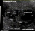
| Stage 13 (week 5) | Stage 22 (week 8) | Stage 23 (week 8) | GIT Abnormalities Ultrasound | ||||||||||||||||
Terms
- allantois - An extraembryonic membrane, endoderm in origin extension from the early hindgut, then cloaca into the connecting stalk of placental animals, connected to the superior end of developing bladder. In reptiles and birds, acts as a reservoir for wastes and mediates gas exchange. In mammals is associated/incorporated with connecting stalk/placental cord fetal-maternal interface.
- amnion - An extraembryonic membrane]ectoderm and extraembryonic mesoderm in origin and forms the innermost fetal membrane, produces amniotic fluid. This fluid-filled sac initially lies above the trilaminar embryonic disc and with embryoic disc folding this sac is drawn ventrally to enclose (cover) the entire embryo, then fetus. The presence of this membane led to the description of reptiles, bird, and mammals as amniotes.
- amniotic fluid - The fluid that fills amniotic cavity totally encloses and cushions the embryo. Amniotic fluid enters both the gastrointestinal and respiratory tract following rupture of the buccopharyngeal membrane. The late fetus swallows amniotic fluid.
- buccal - (Latin, bucca = cheek) A term used to relate to the mouth (oral cavity).
- buccopharyngeal membrane - (oral membrane) (Latin, bucca = cheek) A membrane which forms the external upper membrane limit (cranial end) of the early gastrointestinal tract (GIT). This membrane develops during gastrulation by ectoderm and endoderm without a middle (intervening) layer of mesoderm. The membrane lies at the floor of the ventral depression (stomodeum) where the oral cavity will open and will breakdown to form the initial "oral opening" of the gastrointestinal tract. The equivilent membrane at the lower end of the gastrointestinal tract is the cloacal membrane.
- cloacal membrane - Forms the external lower membrane limit (caudal end) of the early gastrointestinal tract (GIT). This membrane is formed during gastrulation by ectoderm and endoderm without a middle (intervening) layer of mesoderm. The membrane breaks down to form the initial "anal opening" of the gastrointestinal tract.
- coelom - Term used to describe a space. There are extraembryonic and intraembryonic coeloms that form during vertebrate development. The single intraembryonic coelom will form the 3 major body cavities: pleural, pericardial and peritoneal.
- foregut - The first of the three part/division (foregut - midgut - hindgut) of the early forming gastrointestinal tract. The foregut runs from the buccopharyngeal membrane to the midgut and forms all the tract (esophagus and stomach) from the oral cavity to beneath the stomach. In addition, a ventral bifurcation of the foregut will also form the respiratory tract epithelium.
- gastrula - (Greek, gastrula = little stomach) A stage of an animal embryo in which the three germ layers ([E#endoderm|endoderm]/mesoderm/ectoderm) have just formed.
- gastrulation - The process of differentiation forming a gastrula. Term means literally means "to form a gut" but is more in development, as this process converts the bilaminar embryo (epiblast/hypoblast) into the trilaminar embryo ([E#endoderm endoderm]/mesoderm/ectoderm) establishing the 3 germ layers that will form all the future tissues of the entire embryo. This process also establishes the the initial body axes.
- hindgut - The last of the three part/division foregut - midgut - hindgut) of the early forming gastrointestinal tract. The hindgut forms all the tract from the distral transverse colon to the cloacal membrane and extends into the connecting stalk (placental cord) as the allantois. In addition, a ventral of the hindgut will also form the urinary tract (bladder, urethra) epithelium.
- intraembryonic coelom - The "horseshoe-shaped" space (cavity) that forms initially in the third week of development in the lateral plate mesoderm that will eventually form the 3 main body cavities: pericardial, pleural, peritoneal. The intraembryonic coelom communicates transiently with the extraembryonic coelom.
- neuralation - The general term used to describe the early formation of the nervous system. It is often used to describe the early events of differentiation of the central ectoderm region to form the neural plate, then neural groove, then neural tube. The nervous system includes the central nervous system (brain and spinal cord) from the neural tube and the peripheral nervous system (peripheral sensory and sympathetic ganglia) from neural crest. In humans, early neuralation begins in week 3 and continues through week 4.
- neural crest - region of cells at the edge of the neural plate that migrates throughout the embryo and contributes to many different tissues. In the gastrointestinal tract it contributes mainly the enteric nervous system within the wall of the gut responsible for peristalsis and secretion.
- pharynx - uppermost end of gastrointestinal and respiratory tract, in the embryo beginning at the buccopharyngeal membrane and forms a major arched cavity within the phrayngeal arches.
- somitogenesis The process of segmentation of the paraxial mesoderm within the trilaminar embryo body to form pairs of somites, or balls of mesoderm. A somite is added either side of the notochord (axial mesoderm) to form a somite pair. The segmentation does not occur in the head region, and begins cranially (head end) and extends caudally (tailward) adding a somite pair at regular time intervals. The process is sequential and therefore used to stage the age of many different species embryos based upon the number visible somite pairs. In humans, the first somite pair appears at day 20 and adds caudally at 1 somite pair/4 hours (mouse 1 pair/90 min) until on average 44 pairs eventually form.
- splanchnic mesoderm - Gastrointestinal tract (endoderm) associated mesoderm formed by the separation of the lateral plate mesoderm into two separate components by a cavity, the intraembryonic coelom. Splanchnic mesoderm is the embryonic origin of the gastrointestinal tract connective tissue, smooth muscle, blood vessels and contribute to organ development (pancreas, spleen, liver). The intraembryonic coelom will form the three major body cavities including the space surrounding the gut, the peritoneal cavity. The other half of the lateral plate mesoderm (somatic mesoderm) is associated with the ectoderm of the body wall.
- stomodeum - (stomadeum, stomatodeum) A ventral surface depression on the early embryo head surrounding the buccopharyngeal membrane, which lies at the floor of this depression. This surface depression lies between the maxillary and mandibular components of the first pharyngeal arch.
| Lab 5: Introduction | Trilaminar Embryo | Early Embryo | Late Embryo | Fetal | Postnatal | Abnormalities | Online Assessment |
Glossary Links
- Glossary: A | B | C | D | E | F | G | H | I | J | K | L | M | N | O | P | Q | R | S | T | U | V | W | X | Y | Z | Numbers | Symbols | Term Link
Cite this page: Hill, M.A. (2024, May 21) Embryology ANAT2341 Lab 5 - Early Embryo. Retrieved from https://embryology.med.unsw.edu.au/embryology/index.php/ANAT2341_Lab_5_-_Early_Embryo
- © Dr Mark Hill 2024, UNSW Embryology ISBN: 978 0 7334 2609 4 - UNSW CRICOS Provider Code No. 00098G