File:West06.jpg

Original file (308 × 802 pixels, file size: 39 KB, MIME type: image/jpeg)
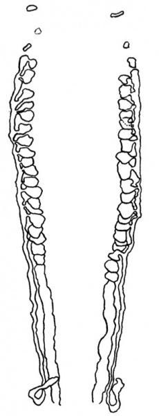
Fig. 6. Mesonephric System
This is composed of a few isolated pronephric rudiments and of a series of mesonephric tubules, or vesicles, which are connected with each other through a longitudinal mesonephric duct that runs caudalwards and just fails to open into the cloaca (Text-fig.6).
The pronephric rudiments are three on each side.
On the left side the first indication of a pronephric tubule occurs as a thickening and invagination of the coelomic epithelium on a level with the middle of the hepatic diverticulum and with about the middle of the 8th somite; the invagination is directed at first dorsalwards and then makes connexion with a small, blind and almost solid, though still hollow, vesicle. The second rudiment is much smaller; it is placed opposite the caudal half of the 9th somite, caudal to the hepatic diverticulum, and it arises in the same manner as the first, but the invagination is directed lateralwards rather than dorsalwards and endsina small, solid clump of cels, the whole structure being much smaller than the first and not extending so far in a dorsal direction. The third rudiment is larger than either the first or second; it is related to about the middle of the 10th somite, and is essentially similar to the first and second tubules and ends in a definitely hollow vesicle.
| Historic Disclaimer - information about historic embryology pages |
|---|
 |
- Links: Fig 1 | Fig 2 | Fig 3 | Fig 4 | Fig 5 | Fig 6 | Fig 7 | Fig 8 | Fig 9 | Fig 10 | Fig 11 | Plate 1 | Plate 1 Fig 1 | Plate 1 Fig 2 | Plate 1 Fig 3 | Plate 1 Fig 4 | Plate 1 Fig 5 | Plate 1 Fig 6
Reference
West CM. A human embryo of twenty-five somites. (1937) J. Anat., 71(2): 169-200.1. PMID 17104635
Cite this page: Hill, M.A. (2024, April 18) Embryology West06.jpg. Retrieved from https://embryology.med.unsw.edu.au/embryology/index.php/File:West06.jpg
- © Dr Mark Hill 2024, UNSW Embryology ISBN: 978 0 7334 2609 4 - UNSW CRICOS Provider Code No. 00098G
File history
Click on a date/time to view the file as it appeared at that time.
| Date/Time | Thumbnail | Dimensions | User | Comment | |
|---|---|---|---|---|---|
| current | 15:21, 28 January 2012 | 308 × 802 (39 KB) | S8600021 (talk | contribs) | ==Fig. 6 == {{Template:West1937}} {{Historic Disclaimer}} {{Historic Papers}} ===Reference=== <pubmed>17104635</pubmed>| [http://www.ncbi.nlm.nih.gov/pmc/articles/PMC1252340 PMC1252340] Category:Carnegie Stage 12 |
You cannot overwrite this file.
File usage
The following page uses this file: