File:West03.jpg

Original file (237 × 1,025 pixels, file size: 25 KB, MIME type: image/jpeg)
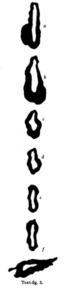
Fig. 3 Outlines of sections of the gut at different levels
The part of the alimentary tract that succeeds the pharynx has been a source of difficulty in interpretation to several observers; in the present specimen it shows, at first, a slit-like lumen and walls of uniform thickness, but, after eight sections, there appears a rather spherical outgrowth on the right side (Text-fig. 3a) and, after a few more sections, there is a corresponding dilatation on the left; the difference in level of the two outgrowths is due to a slight obliquity of the sections through this part of the gut. These two out-growths, which I will call the cranial swellings, arise from the ventral part of the gut,whereas the dorsal part of the gut has a slit-like lumen and is separated from the ventral expanded part by a slight constriction; beyond this cranial swelling the gut shows a lumen which is suddenly much reduced in ventro- dorsal diameter, being the continuation merely of the slit-like dorsal part of the more cranial portion of the gut; farther on the gut has a pear-shaped lumen, wide ventrally where it forms what I will call the caudal swelling (Text-fig.3d), and slit-like dorsally, and it becomes continuous, in the septum transversum with the part of the gut from which the liver arises (Text-fig. 3f).
The cranial swelling shows a slight indication of division into two; its walls are much thickened; it is to the cranial side of the septum transversum; it lies dorsal to the sinus venosus and atrium, just where these two cavities communicate; the swellings is greater towards the left side than to the right, and on the left side it grows towards the pericardial cavity and on the right towards the pleural passage. The caudal swelling is much smaller; it shows no evidence of duplicity; it is close to the hepatic diverticulum, and it shows no thickening of the ventral wall, which is, indeed, rather thinner than the dorsal wall. I show (Text-fig.3) a series of outlines of sections of the gut at different levels,which may be compared with similar figures by van den Broek, and by Girgis and others, and from which it will be seen that I am in disagreement with van den Broek and Girgis, both of whom show what they describe as lung, in the same section as the liver.
| Historic Disclaimer - information about historic embryology pages |
|---|
 |
- Links: Fig 1 | Fig 2 | Fig 3 | Fig 4 | Fig 5 | Fig 6 | Fig 7 | Fig 8 | Fig 9 | Fig 10 | Fig 11 | Plate 1 | Plate 1 Fig 1 | Plate 1 Fig 2 | Plate 1 Fig 3 | Plate 1 Fig 4 | Plate 1 Fig 5 | Plate 1 Fig 6
Reference
West CM. A human embryo of twenty-five somites. (1937) J. Anat., 71(2): 169-200.1. PMID 17104635
Cite this page: Hill, M.A. (2024, April 20) Embryology West03.jpg. Retrieved from https://embryology.med.unsw.edu.au/embryology/index.php/File:West03.jpg
- © Dr Mark Hill 2024, UNSW Embryology ISBN: 978 0 7334 2609 4 - UNSW CRICOS Provider Code No. 00098G
File history
Click on a date/time to view the file as it appeared at that time.
| Date/Time | Thumbnail | Dimensions | User | Comment | |
|---|---|---|---|---|---|
| current | 15:20, 28 January 2012 | 237 × 1,025 (25 KB) | S8600021 (talk | contribs) | ==Fig. 3 == {{Template:West1937}} {{Historic Disclaimer}} {{Historic Papers}} ===Reference=== <pubmed>17104635</pubmed>| [http://www.ncbi.nlm.nih.gov/pmc/articles/PMC1252340 PMC1252340] Category:Carnegie Stage 12 |
You cannot overwrite this file.
File usage
The following page uses this file: