File:Waterston05.jpg

Original file (414 × 678 pixels, file size: 81 KB, MIME type: image/jpeg)
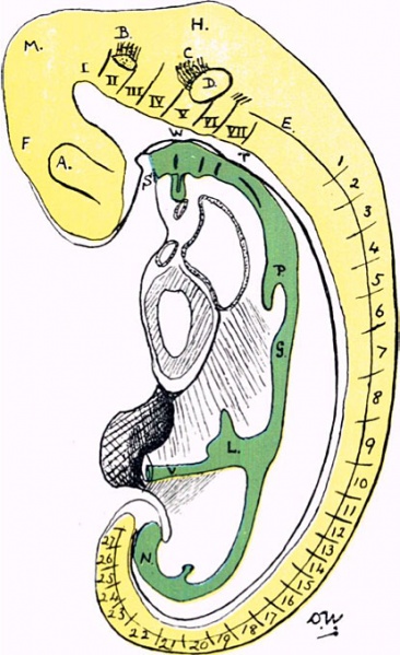
Fig. 5. Lincar reconstruction of nervous system and alimentary canal
Lincar reconstruction of nervous system, alimentary canal, etc. The figures 1-27 indicate somites, I.-VII. neuromeres.
Legend
- A - optic diverticulum
- B - trigeminal nerve and ganglion rudiment
- C - acustico- facial nerve complex
- D - otic vesicle
- E - neural crest
- F - forebrain
- G - stomach
- H - hindbrain
- L - liver-bud
- M - midbrain
- N - cloaca
- P - albove lung-bud
- S - stomatodeum
- T and W - notochord in contact with pharyngeal wall
Carnegie Staging Comparison: A 27 somite stage embryo would be similar to a Carnegie stage 12 (26 - 30 days), caudal neuropore closes, Somite Number 21-29.
- 27 Somite Paper: Fig 1 | Fig 2 | Fig 3 | Fig 4 | Fig 5 | Fig 6 | Fig 7 | Fig 8 | Fig 9 | Fig 10 | Fig 11 | Fig 12 | Fig 13 | Fig 14 | Fig 15 | Fig 16 | Fig 17 | Fig 18 | Fig 19 | Fig 20 | Carnegie stage 12
| Historic Disclaimer - information about historic embryology pages |
|---|
 |
- Historic Paper Links: 13-14 Somites | 22 Somites | 23 Somites | 25 Somites | 27 Somites | Mall Human Embryo Collection | Embryology History | Carnegie stage 11 | Carnegie stage 12 | Journal of Anatomy | Embryonic Development | Category:Historic Embryology
Reference
Waterston D. A human embryo of twenty-seven pairs of somites, embedded in decidua. (1914) J Anat Physiol., 49(1): 90-118 PMID 17233016
Cite this page: Hill, M.A. (2024, April 16) Embryology Waterston05.jpg. Retrieved from https://embryology.med.unsw.edu.au/embryology/index.php/File:Waterston05.jpg
- © Dr Mark Hill 2024, UNSW Embryology ISBN: 978 0 7334 2609 4 - UNSW CRICOS Provider Code No. 00098G
File history
Click on a date/time to view the file as it appeared at that time.
| Date/Time | Thumbnail | Dimensions | User | Comment | |
|---|---|---|---|---|---|
| current | 12:51, 27 January 2012 | | 414 × 678 (81 KB) | S8600021 (talk | contribs) | <pubmed>17233016</pubmed>| [http://www.ncbi.nlm.nih.gov/pmc/articles/PMC1288995 PMC1288995] ===Historic Embryology=== This is a slightly edited version of the original 1914 paper published in Journal of Anatomy and Physiology. The full paper is still av |
You cannot overwrite this file.