File:Uterine tube histology.jpg
Original file (1,280 × 1,024 pixels, file size: 568 KB, MIME type: image/jpeg)
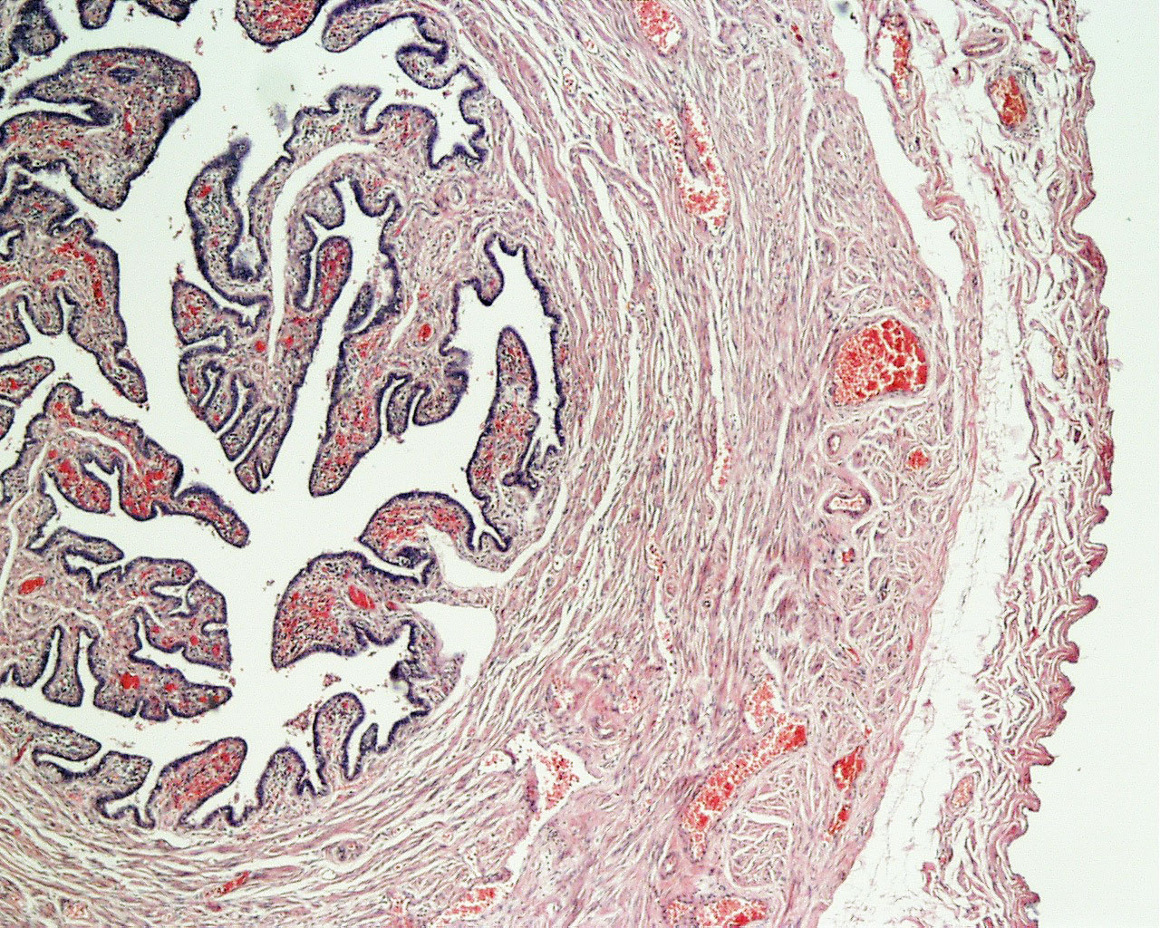
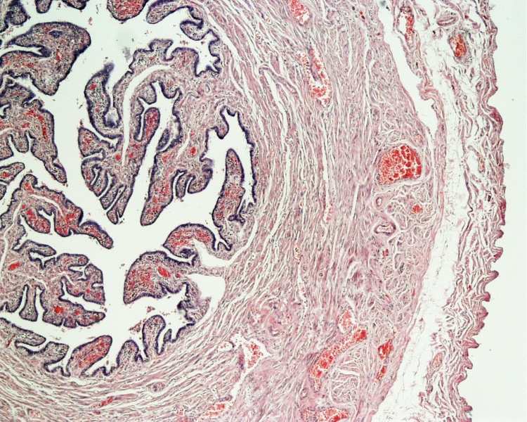
Uterine Tube Histology
Uterine Tube (oviduct, Fallopian tube) functions as a conduit for the oocyte, from the ovaries to the uterus. Histologically, the oviduct consists of a mucosa and a muscularis. The peritoneal surface of the oviduct is lined by a serosa and subjacent connective tissue.
The mucosa
- formed by a ciliated and secretory epithelium resting on a very cellular lamina propria.
- The number of ciliated cells and secretory cells varies along the oviduct.
- Secretory activity varies during the menstrual cycle, and resting secretory cells are also referred to as peg-cells.
- Some of the secreted substances are thought to nourish the oocyte and the very early embryo.
The muscularis
- inner circular muscle layer and an outer longitudinal layer.
- An inner longitudinal layer is present in the isthmus and the intramural part of the oviduct.
- Peristaltic muscle action seems to be more important for the transport of sperm and oocyte than the action of the cilia.
Tube Anatomical Four Subdivisions
- Infundibulum is the funnel-shaped (up to 10 mm in diameter) end of the oviduct. Finger-like extensions of its margins, the fimbriae, are closely applied to the ovary. Ciliated cells are frequent. Their cilia beat in the direction of the ampulla of the oviduct.
- Ampulla - Mucosal folds, or plicae, and secondary folds which arise from the plicae divide the lumen of the ampulla into a very complex shape.. Fertilization usually takes place in the ampulla.
- Isthmus is the narrowest portion (2-3 mm in diameter) of the parts of the oviduct located in the peritoneal cavity. Mucosal folds are less complex and the muscularis is thick. An inner, longitudinal layer of muscle is present in the isthmus.
- Intramural part of the oviduct, which penetrates the wall of the uterus body. An inner, longitudinal layer of muscle is present.
H&E stain
Links: Histology | Histology Stains | Blue Histology images copyright Lutz Slomianka 1998-2009. The literary and artistic works on the original Blue Histology website may be reproduced, adapted, published and distributed for non-commercial purposes. See also the page Histology Stains.
Cite this page: Hill, M.A. (2024, April 25) Embryology Uterine tube histology.jpg. Retrieved from https://embryology.med.unsw.edu.au/embryology/index.php/File:Uterine_tube_histology.jpg
- © Dr Mark Hill 2024, UNSW Embryology ISBN: 978 0 7334 2609 4 - UNSW CRICOS Provider Code No. 00098G
http://www.lab.anhb.uwa.edu.au/mb140/CorePages/FemaleRepro/femalerepro.htm#Uterus
Odu04he.jpg
File history
Click on a date/time to view the file as it appeared at that time.
| Date/Time | Thumbnail | Dimensions | User | Comment | |
|---|---|---|---|---|---|
| current | 22:57, 22 April 2010 | | 1,280 × 1,024 (568 KB) | S8600021 (talk | contribs) | Uterine Tube (oviduct, Fallopian tube) The oviduct functions as a conduit for the oocyte, from the ovaries to the uterus. Histologically, the oviduct consists of a mucosa and a muscularis. The peritoneal surface of the oviduct is lined by a serosa and su |
You cannot overwrite this file.
File usage
The following 2 pages use this file: