File:Upper auricular detachment 01.jpg
From Embryology
No higher resolution available.
Upper_auricular_detachment_01.jpg (412 × 600 pixels, file size: 43 KB, MIME type: image/jpeg)
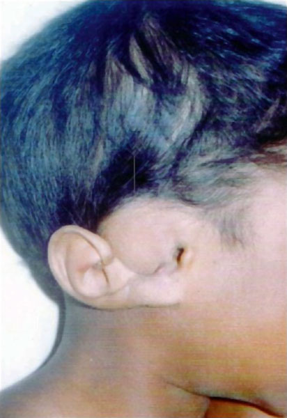
Hearing Development - Upper Auricular Detachment
Profile view of head showing detached external ear, on right side. Upper auricle attached to temple with ear lobule only on right side.
Reference
<pubmed>20368874</pubmed>| PMC2845381 | Indian J Plast Surg.
Copyright
http://creativecommons.org/licenses/by-nc-sa/3.0/
Original file name: Figure 2. http://www.ijps.org/viewimage.asp?img=ijps_2009_42_2_265_59298_u2.jpg (original image resized and cropped)
File history
Click on a date/time to view the file as it appeared at that time.
| Date/Time | Thumbnail | Dimensions | User | Comment | |
|---|---|---|---|---|---|
| current | 11:41, 17 November 2010 | | 412 × 600 (43 KB) | S8600021 (talk | contribs) | ==Hearing Development - Upper auricular detachment== Profile view of head showing detached external ear, on right side. Upper auricle attached to temple with ear lobule only on right side. Original file name: Figure 2. http://www.ijps.org/viewimage.asp? |
You cannot overwrite this file.
File usage
The following page uses this file: