File:Thyroid-agenesis of isthmus.jpg
From Embryology
Size of this preview: 499 × 600 pixels. Other resolution: 574 × 690 pixels.
Original file (574 × 690 pixels, file size: 97 KB, MIME type: image/jpeg)
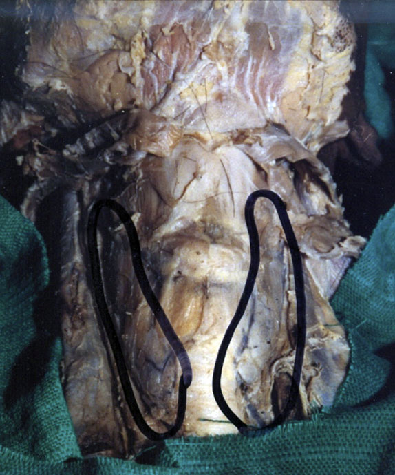
Agenesis of isthmus of thyroid gland
- Two lateral lobes (black outlines) lying independently on either side of the trachea connected by a thin layer of pre-tracheal fascia.
- The normal isthmus measures about 1.25 cm transversely as well as vertically and is located anterior to the second and third tracheal cartilages.
Original file name: Figure 3 http://www.ncbi.nlm.nih.gov/pmc/articles/PMC2827060/figure/F3/
Reference
<pubmed>20181171</pubmed>| PMC2827060
This is an Open Access article distributed under the terms of the Creative Commons Attribution License (http://creativecommons.org/licenses/by/3.0), which permits unrestricted use, distribution, and reproduction in any medium, provided the original work is properly cited.
File history
Click on a date/time to view the file as it appeared at that time.
| Date/Time | Thumbnail | Dimensions | User | Comment | |
|---|---|---|---|---|---|
| current | 23:27, 5 October 2010 | | 574 × 690 (97 KB) | S8600021 (talk | contribs) | ==Agenesis of isthmus of thyroid gland== Two lateral lobes (black outlines) lying independently on either side of the trachea connected by a thin layer of pre-tracheal fascia. Original file name: Figure 3 http://www.ncbi.nlm.nih.gov/pmc/articles/PMC282 |
You cannot overwrite this file.
File usage
The following page uses this file: