File:T2 lymphocyte EM14.jpg
Original file (728 × 771 pixels, file size: 156 KB, MIME type: image/jpeg)
T Lymphocyte Electron Micrograph
Effector T cell has relatively little cytoplasmic rough endoplasmic reticulum (RER) but is filled with free ribosomes.
Cytotoxic T lymphocytes (CTLs) (killer T cells) - directly attack other cells carrying certain foreign or abnormal molecules on their surfaces. CTLs are especially useful for attacking viruses because viruses often hide from other parts of the immune system while they grow inside infected cells. CTLs recognize small fragments of these viruses peeking out from the cell membrane and launch an attack to kill the infected cell.
Helper T cells (Th cells) - coordinate immune responses by communicating with other cells. Some stimulate nearby B cells to produce antibodies, others call in microbe-gobbling cells called phagocytes, and still others activate other T cells.
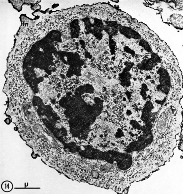
Fig. 14. Lymph node cells (4 days after immunization with DBA/2 mastocytoma) labeled with aBA0 followed by SaRIg-Pox. This cell classified as T2 lymphocyte is characterized by its large content in polyribosomes, often forming "rosettes," and by an enveloping filamentous network (fn) , appearing at this magnification as a peripheral greyish area.
X 18,500.
Note this T2/T3 terminology may now be historic.
- Lymphocyte EM Images: T and B Lymphocytes 1 TEM | T and B Lymphocytes 2 TEM | T Lymphocyte SEM | B lymphocyte 1 TEM | B lymphocyte 2 TEM | B lymphocyte 3 TEM | Plasma Cell TEM | T2 Lymphocyte 1 TEM | T2 Lymphocyte 2 TEM | lymphocyte rosettes | T lymphocyte 1 | T lymphocyte 2 | T lymphocyte 3 | T lymphocyte 4 | T lymphocyte 5 | T lymphocyte 6 | B lymphocyte | B lymphocytes TEM | Immune System Development | Blood
Reference
Matter A, Lisowska-Bernstein B, Ryser JE, Lamelin JP & Vassalli P. (1972). Mouse thymus-independent and thymus-derived lymphoid cells. II. Ultrastructural studies. J. Exp. Med. , 136, 1008-30. PMID: 4563148
Copyright
Rockefeller University Press - Copyright Policy This article is distributed under the terms of an Attribution–Noncommercial–Share Alike–No Mirror Sites license for the first six months after the publication date (see http://www.jcb.org/misc/terms.shtml). After six months it is available under a Creative Commons License (Attribution–Noncommercial–Share Alike 4.0 Unported license, as described at https://creativecommons.org/licenses/by-nc-sa/4.0/ ). (More? Help:Copyright Tutorial)
Cite this page: Hill, M.A. (2024, April 25) Embryology T2 lymphocyte EM14.jpg. Retrieved from https://embryology.med.unsw.edu.au/embryology/index.php/File:T2_lymphocyte_EM14.jpg
- © Dr Mark Hill 2024, UNSW Embryology ISBN: 978 0 7334 2609 4 - UNSW CRICOS Provider Code No. 00098G
File history
Click on a date/time to view the file as it appeared at that time.
| Date/Time | Thumbnail | Dimensions | User | Comment | |
|---|---|---|---|---|---|
| current | 16:30, 22 February 2012 | | 728 × 771 (156 KB) | Z8600021 (talk | contribs) | ==T2 lymphocyte Electron Micrograph== Fig. 14. Lymph node cells (4 days after immunization with DBA/2 mastocytoma) labeled with aBA0 followed by SaRIg-Pox. This cell classified as T2 lymphocyte is characterized by its large content in polyribosomes, ofte |
You cannot overwrite this file.
File usage
The following 4 pages use this file: