File:Streeter1922-fig14.jpg

Original file (617 × 672 pixels, file size: 70 KB, MIME type: image/jpeg)
Fig. 14. Human Embryo 11mm long
Human Embryo No. 1767, 11 mm
In embryos 4 to 6 mm. long the mandibular and hyoid bars are each subdivided by a transverse groove into a dorsal and a ventral part, as can be seen in figures 13 and 14 (plate 2); also figure 9 (plate 1). These are not to be confused with the branchial hillocks. The significance of the subdivision of these bars has never been determined; we shall see, however, that the closure mechanism is derived from the ventral portions, while the articular and sound-collecting mechanisms are derived from the dorsal portions. His (1882), in describing the mandibular bar in young embryos, mentions the existence of a root part (Wurzelstiick) as distinguished from the more ventral portion, which he describes as divided longitudinally into a lip ridge (Lippenwulst) and a mental ridge (Kinnwulst). Careful examination of figures 10 and 11 (plate 1) will show that the ventral part of the mandibular arch is roughly subdivided into two ridges, somewhat as described by His. These ridges do not, however, correspond exactly to the eventual chin and lip, as His first thought. The more anterior one (lip-ridge) in reality gives origin to the greater part of the jaw, the Up being a much later derivative of it. The more posterior ridge (Kinnwulst) corresponds to the soft parts beneath the jaw.
Plate 2. Drawings of human embryos, showing the region of the first branchial cleft and its transformation into a fossa angularis.
Coincident with this transformation the mesenchyme of the hyoid and mandibular bars undergoes proliferation and becomes condensed to form the primordium of the auricle. Foci of more active proliferation show on the surface as branchial hillocks. Specimens are from the Carnegie Collection.
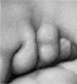
Fig. 13. Embryo No. 1380, 5 mm
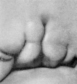
Fig. 14. Embryo No. 1767, 11 mm
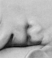
Fig. 15. Embryo No. 1461, 10 mm
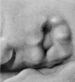
Fig. 16. Embryo No. 562, 13 mm

Fig. 17. Embryo No. 1232, 14 mm
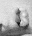
Fig. 18. Embryo No. 475, 15 mm


- In-text Figures: Figure 1 and 2 | Figure 3 and 4 | Figure 5 | Figure 6 and 7 | Figure 8 | Text | Glossary
- Plates: Plate 1 | Plate 2 | Plate 3 | Plate 4 | Plate 5 | Plate 6 | Plates | Glossary
- Figures: 1. Auricle cartilage | 2. External ear | 3. Agnathia | 4. Agnathia+cyclopia | 6. Auricular cartilage embryo 21, 32 and 43 mm | 7. Auricular cartilage 50 mm fetus | 9. Embryo 6 mm | 10. Embryo 12 mm | 11. Embryo 14 mm | 12. Embryo 18 mm | 13. Embryo 1380, 5 mm | 14. Embryo 1767, 11 mm | 15. Embryo 1461, 10 mm | 16. Embryo 562, 13 mm | 17. Embryo 1232, 14 mm | 18. Embryo 475, 15 mm | 19. Embryo 899, 13 mm | 20. Embryo 434, 15 mm | 21. Embryo 492, 16.8 mm | 22. Embryo 576, 17 mm | 23. Embryo 547, 18 mm | 24. Embryo 955, 17 mm | 25. Embryo 1584, 18 mm | 26. Embryo 1134e, 21.3 mm | 27. Embryo 1358b, 33.2 mm | 28. Embryo 1535, 28 mm | 29. Embryo 2163, 36 mm | 30. Embryo 1980, 37 mm | 31. Embryo 1840a, 38.5 mm | 32. Embryo 2075, 40 mm | 33. Embryo 2144, 45.5 mm | 34. Embryo 642, 49 mm | 35. Embryo 2170, 50 mm | 36. Embryo 2095, 52 mm | 37. Embryo 2095, 52 mm | 38. Embryo 2066, 53 mm | 39. Embryo 2079, 56.5 mm. | 40. Embryo 1561, 57 mm | 41. Embryo 218, 62.5 mm. (R.) | 42. Embryo 1724, 66.2 mm | 43. Embryo 2328, 65 mm | 44. Embryo 2118, 69 mm | 45. Embryo 981, 85 mm | 46. Embryo 1845, 87 mm | 47. Embryo 1449, 87.3 mm | 48. Embryo 2003, 103.5 mm | 49. Embryo 1858, 100 mm | 50. Embryo 2274, 113 mm | 51. Embryo 2185, 113.5 mm. | 52. Embryo 9526, 114 mm. | 53. Embryo 1811, 114 mm | 54. Embryo 1716, 119 mm. Fig. 59. 1742, 191.2 mm | 55. Embryo 19576, 119 mm. | 56. Embryo 1782, 135.6 mm | 57. Embryo 1702, 150 mm | 58. Embryo 1708, 154 mm | 59. Embryo 1742, 191.2 mm | Figures
- Related Notes: Outer Ear Development | Carnegie Contributions to Embryology
Reference
Streeter GL. Development of the auricle in the human embryo. (1922) Carnegie Instn. Wash. Publ. 277, Contrib. Embryol., 14: 111-138.
Cite this page: Hill, M.A. (2024, April 24) Embryology Streeter1922-fig14.jpg. Retrieved from https://embryology.med.unsw.edu.au/embryology/index.php/File:Streeter1922-fig14.jpg
- © Dr Mark Hill 2024, UNSW Embryology ISBN: 978 0 7334 2609 4 - UNSW CRICOS Provider Code No. 00098G
| Historic Disclaimer - information about historic embryology pages |
|---|
 |
File history
Click on a date/time to view the file as it appeared at that time.
| Date/Time | Thumbnail | Dimensions | User | Comment | |
|---|---|---|---|---|---|
| current | 12:56, 27 January 2013 | | 617 × 672 (70 KB) | Z8600021 (talk | contribs) | ==Fig. 14. Human Embryo 11mm long== Plate 2. Drawings of human embryos, showing the region of the first branchial cleft and its transformation into a fossa angularis. Coincident with this transformation the mesenchyme of the hyoid and mandibular bars un |
You cannot overwrite this file.
File usage
The following 8 pages use this file: