File:Streeter1921 fig27.jpg
From Embryology
Size of this preview: 569 × 600 pixels. Other resolution: 949 × 1,000 pixels.
Original file (949 × 1,000 pixels, file size: 138 KB, MIME type: image/jpeg)
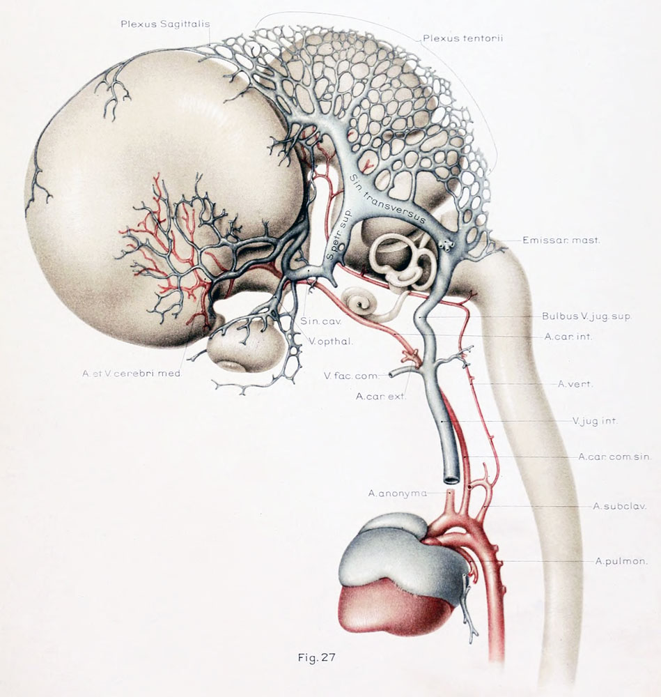
Figure 27. Left profile view of a wax-plate reconstruction of the blood-vessels of the brain in a human embryo 43 mm long
Carnegie Collection Embryo No. 886
Enlarged 8 diameters. (original print version)
- The vascular architecture is at this stage beginning to approximate the adult condition, although the whole tentorial region retains its embryonic character, due to which the marked subsequent migration of the transverse sinus is possible.
- It will be noted that the sigmoid portion of the sinus is fairly well established.
--Mark Hill 09:39, 17 February 2011 (EST) Post-Embryonic based upon CRL. Figure same as cropped Plate 5.
- 1921 Human Brain Vascular: Fig 1 | Fig 2 | Fig 3 | Fig 4 | Fig 5 | Fig 6 | Fig 7-9 | Fig 10 | Fig 11 | Fig 12 |Fig 13 | Fig 14 | Fig 15 | Fig 16 | Fig 17 | Fig 18 | Fig 19 | Fig 20 | Fig 21 | Fig 22 | Fig 23 | Fig 24 | Fig 25 | Fig 26 | Fig 27 | Plate 1 - embryos 4 mm to birth | Plate 2 - embryo 4 mm | Plate 3 - embryo 11.5 mm | Plate 4 - embryo 21 mm | Plate 5 - embryo 43 mm | Carnegie No.24 | George Streeter
| Historic Disclaimer - information about historic embryology pages |
|---|
 |
Reference
Streeter GL. The developmental alterations in the vascular system of the brain of the human embryo. (1921) Contrib. Embryol., Carnegie Inst. Wash. 8:7-38.
Cite this page: Hill, M.A. (2024, April 20) Embryology Streeter1921 fig27.jpg. Retrieved from https://embryology.med.unsw.edu.au/embryology/index.php/File:Streeter1921_fig27.jpg
- © Dr Mark Hill 2024, UNSW Embryology ISBN: 978 0 7334 2609 4 - UNSW CRICOS Provider Code No. 00098G
File history
Click on a date/time to view the file as it appeared at that time.
| Date/Time | Thumbnail | Dimensions | User | Comment | |
|---|---|---|---|---|---|
| current | 16:34, 21 April 2012 | | 949 × 1,000 (138 KB) | Z8600021 (talk | contribs) | ==Figure 27. Left profile view of a wax-plate reconstruction of the blood-vessels of the brain in a human embryo 43 mm long == Carnegie Collection Embryo No. 886 Enlarged 8 diameters. (original print version) * The vascular architecture is at this stag |
You cannot overwrite this file.
File usage
There are no pages that use this file.