File:Streeter1920chart5.jpg
From Embryology

Size of this preview: 800 × 502 pixels. Other resolution: 1,000 × 627 pixels.
Original file (1,000 × 627 pixels, file size: 74 KB, MIME type: image/jpeg)
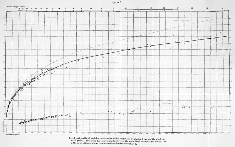
Chart 5. Field and curve of mean menstrual age for specimens in the Carnegie Collection having menstrual histories
The circles placed at the 32d, 36th, and 40th weeks are taken from Zangemeister's (1911) data and are used to complete the curve to the termination of pregnancy.
| Historic Disclaimer - information about historic embryology pages |
|---|
 |
- Embryo Size Links: Fig. 1 Fetus grades | Fig. 2 Measuring Instrument | Table 1 sitting height and weight | Table 2 Weight | Table 3 variation height and weight | Table 4 Foot length | Table 5 Head modulus | Table 6 embryos less than 10 grams | Table 7 Page 1 embryos more than 10 grams | Table 7 Page 2 embryos more than 10 grams | Table 7 Page 3 embryos more than 10 grams | Table 7 Page 4 embryos more than 10 grams | Table 7 Page 5 embryos more than 10 grams | Table 7 Page 6 embryos more than 10 grams | Table 7 Page 7 embryos more than 10 grams | Table 7 Page 8 embryos more than 10 grams | Table 7 Page 9 embryos more than 10 grams | Table 7 Page 10embryos more than 10 grams | Chart 1 crown-rump length | Chart 2 curve of growth | Chart 3 Foot length and head modulus | Chart 4 Carnegie Collection over 200 grams | Chart 5 weight and head modulus | Contributions to Embryology
Reference
Streeter GL. Weight, sitting height, head size, foot length, and menstrual age of the human embryo. (1920) Contrib. Embryol., Carnegie Inst. Wash. Publ. , : 143-170.
Cite this page: Hill, M.A. (2024, April 19) Embryology Streeter1920chart5.jpg. Retrieved from https://embryology.med.unsw.edu.au/embryology/index.php/File:Streeter1920chart5.jpg
- © Dr Mark Hill 2024, UNSW Embryology ISBN: 978 0 7334 2609 4 - UNSW CRICOS Provider Code No. 00098G
| Historic Disclaimer - information about historic embryology pages |
|---|
|
File history
Click on a date/time to view the file as it appeared at that time.
| Date/Time | Thumbnail | Dimensions | User | Comment | |
|---|---|---|---|---|---|
| current | 15:47, 30 January 2012 | | 1,000 × 627 (74 KB) | S8600021 (talk | contribs) | {{Streeter1920}} {{Historic Disclaimer}} |
You cannot overwrite this file.
File usage
The following page uses this file: