File:Streeter1920 02.jpg
Original file (763 × 1,000 pixels, file size: 56 KB, MIME type: image/jpeg)
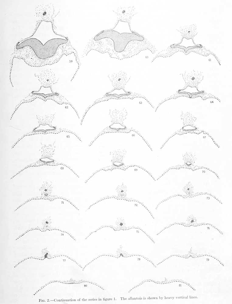
Fig. 2. Outline serial drawings showing the form of the embryonic plate and its relations to the yolk-sac and the amniotic cavity
Outline serial drawings showing the form of the embryonic plate and its relations to the yolk-sac and the amniotic cavity. The numbers refer to the section number. Enlargement 50 diameters.
Section 59: (Compare fig. 14, plate 3.) The body-stalk consists of two portions — a round, more condensed poition surrounding the allantoic duct, and outside of this a triangular area of looser mesodcniial tissue which extends up to unite with the chorionic membrane. In the more condensed portion several endothelial spaces can be seen. The allantoic duct contains a lumen. The formation of endodemi, mesoderm, and ectodenn is similar to that in the last section. What appear to be beginning bloodislands can be seen in the ventral part of the yolk-sac.
Section 60: This section was cut 40^ thick, otherwise it is much the same as section 59. The body-stalk appears as a very condensed mass and at its center can be seen the allantoic duct with a narrow lumen. The stalk is partly covered with a distinct mesodemial membrane. The amniotic cavity fits close against its ventral wall and is considerably contracted, due to the fact that the lateral intermediate plate lies against the main embryonic plate, thereby reducing the width of the cavity by nearly onehalf. ()wing to the thickness of the section the details of the fusion between the ectoderm, mesoderm, and entloderm can not be made out. As in the previous sections, however, there is no indication of a ni'urcntcric canal.
Section 61: The transition from sections 60 to 61 appears to be very marked, but is due merely to the thickness of the preceding section, which conceals the change in form which the embryo umlergoes at this point. By focusing up and down through the section one can recognize the change from a broad tangential section through the embryonic plate to a thin, narrower, transverse section, sutlicient to account for the transition between these two sections. In section 61 the compact portion of the bodystalk is sejiarated from the chorionic membrane by the Icioser mesodemial tissue referred to in the descri])tion of previous sections. The attachment is maintained only by loose siraiuls of mesodennal cells. The allantoic stalk is very much constricted in this section and consists of only a few ceils which are much less compact than in the adjacent sections. Several endothelial spaces can be recognized in the compact portion of the body-stalk. A distinct mesodermal membrane incloses the ventral half of the stalk on each side, spreading over the amnion, whence it continues down over the yolk-sac, constituting the outer of the two layers of the wall of the latter. In the dorsal half of the wall it is distinct from the endodermal layer; ventral to this the two closely fuse and can no longer be distinguished as separate layers. The amniotic ectoderm fits closely against the round ventral surface of the body-stalk and laterally extends downward to a point where it becomes continuous with the transitional portion of the embryonic plate. The embryonic plate proper shows a sharply cut primitive groove, at which point the plate fuses with the endoderm of the yolk-sac. Whether any mesoderm is interposed in this section can not be definitelj' determined. Lateral to this point there is a considerable amount of mesodermal tissue intervening between the embryonic plate and the yolk-sac, being everywhere closely adherent to the former. It is connected with the yolk-sac by a few slender strands which mark off a series of clear, round spaces, the most lateral of which is continuous with a cleft separating the mesoderm and endodenn from the dorsal portion of the yolksac. No endothelial spaces seem to be present in this region.
Section 63: The compact portion of the bodystalk is still farther removed from the chorionic membrane than in the previous section. The allantoic stalk is now somewhat larger, but no lumen can be recognized. A small, endothelial like ring of cells lies free in the exoccelom lateral to the mesodennal membrane covering the bodystalk. This section passes through the amniotic cavity in a transveree direction favorable for showing the structure of its ectodennal walls. Three distinct regions can be made out — the flattened amniotic ectoderm, the transitional lateral embryonic plate (consisting of one layer of cylindrical cells), and the embryonic plate proper (consisting of two or three layers of cylindrical epithehal cells). At the primitive groove the ectodenn is in contact with the endoderm of the yolk-sac. Lateral to this point there is a considerable amount of mesodermal tissue which has the appearance of flowing out from the lateral portions of the embryonic plate. Strands from this mesoderm extend out to the endoderm of the yolk-sac, outhning spjices similar to those described in the last section. The wall of the yolk-sac in its dorsal half consists of two distinct layers — mesoderm and endoderm. More vent rally the two layers fuse, and in the extreme ventral pole there would appear, in places, to be only one lajer, endoilerm. At a few points on the ventral portion of the yolksac there may be seen clusters of 3 or 4 nuclei, which possibly represent beginning angioblasts.
Sections 63 and 64: In the body-stalk and in the loose mesodermal tissue between it and the chorionic membrane are several endothelium lined spaces. In section 64, in the center of the body-stalk, can be seen a well-defined allantoic stalk containing a lumen. The form and structure of the amniotic cavity and its walls are much the same as in the previous section. The relation of the embrj-onic plate to the yolk-sac is very intimate in the region of the primitive groove. The embryonic plate shows the presence of numerous division figures. The mesoderm intervening between the plate and the endoderm of the yolk-sac is closely adherent to the former. A more detailed drawing of this section is shown in figure 13, plate 3.
Sections 65 and 66: In the loose tissue between the body-stalk and the chorionic membrane is an elongated space, in the lumen of which are a few cells. This is the largest space thus far encountered. The lateral surfaces of the compact portion of the body-stalk are entirely walled in by a mesodennal membrane. In the region of these sections the allantoic stalk is interrupted; at a point where it should be present one sees only the same mesodermal tissue found in other parts of the body-stalk. The amniotic cavity is rapidly contracting; its apex remains flattened in conformity to the ventral contour of the bodystalk. The embryonic plate is much narrower and the primitive groove is still sharply cut. The ectodenn at this point is not so closely adherent to the endoderm of the yolk-sac as in the previous sections. The ventral portion of the wall of the yolk-sac is very much thinned out, and one can not be sure that it consists of more than one layer. In the dorsal portions, however, an outer mesodermal membrane is sharply set off from the endoderm.
Sections 67 and 68: In the body-stalk is an open space in the area where one would expect to find the allantoic stalk, but otherwise there is no trace of that structure. The amniotic cavity is further contracted (see fig. 12, plate 2). The embryonic plate forming its floor still shows the characteristic outlines of a primitive groove at the center. It is thinner than in the preceding sections, consisting of one to two layers of cells, and the middle is no longer in contact with the endodenu of the yolk-sac. The mesoderm between the embryonic plate and the yolk-sac is predominantly adherent to the former, being separated from the latter by a series of spaces similar to those described in the previous sections. In the mesoderm of this region there is no evidence of blood-vessel fonnation. To the left of the loose tissue, in section 68, intervening between the body-stalk and the chorionic membrane is a group of mesodennal cells which take part in the fonnation of a structure that will be followed in the next eleven sections. There is so trace of the allantoic stalk. The primitive groove can still he recognized. The ectoderm at this point, however, is not in contact with the endodcnn of the yolk-sac. As in previous sections, the ventral portion of the yolk-sac shows very little evidence of being blood-vessel formation.
Section 69: The amniotic cavity is more contracted and still shows the presence of a primitive groove. The relation of the embryonic plate to the mesoderm intervening between it .'uui the yolk-sac is less closely maintained than in the foregoing sections. In the ventral portion of the yolk-sac is a distinct group of angioblasts, consisting of a strand of about 12 cells. On each side of the strand the wall of the yolk-sac is very thin.
Section 70: The group of mesodermal cells referred to in the last section can now be recognized as arranged in the form of a membrane, cut tangentially. The compact portion of the botiy-stalk is much smallerj and the only evidence it shows of an allantoic stalk is a doubtful open space. The amniotic cavity is now very small; its ventral floor still ha.s the characteristics of the embryonic plate in contra.st to the thin amniotic ectoderm of its roof, as can be seen in figure 11, plate 2. Several angiogenetic areas can be recognized in the ventral portion of the yolk-sac.
Section 71: The body-stalk is now somewhat detached from the loose mesodermal tissue intervening between it and the chorionic membrane. It contains the beginning of the main portion of the allantoic stalk and the tip of the amniotic cavity, the floor of which consists of a small group of ectodermal cells projecting ventrally. The ventral portion of the yolk-sac shows a continuation of the angiogenesis referred to in the last section.
Section 72: Dorsal to the body-stalk can be seen two separate masses, each of which has an average diameter of about the thickness of the chorionic membrane. The one to the left is a contiimation of the mesodcmiic membrane seen in the previous section, and here it can be seen that the mesoderm incloses a solid mass of ectodermal cells of two kinds; a dorsal, paler group, and a ventral, deeply staining group, the two being sharply marked off from one another. The other mass is somewhat less compact and consists partly of mesodenn and partly of cells whose form is better seen in section 73. In the center of the abdominal stalk is the allantoic stalk, sharply marked off and with a clearly defined lumen. Ventral to this is the tip of the amniotic cavity, whose walls are still dilferontiatcd in the dorsal amniotic ectodenii and the ventral embryonic jjlate. The yolk-sac can now be seen at about its greatest diameter, and is spherical in outline. Its dorsal third is composed of two separate layers, mesoderm and ectoderm. In the ventral two-thirds the layers are so intimately fused that they can not be distinguished; at the extreme ventral pole they have the appearance of a single layer, although the existence of angioblasts in this region indicates the presence of mesodermal elements. One group of angioblasts consists of a round, compact clmnp of 5 nuclei. The largest group gives the appearance of an elongated oval endothelial space, compactly filled with about 15 nuclei.
Section 73: The character of the two small masses seen in the previous sections, in the space intervening between the body-stalk and the chorionic membrane, can now be clearly made out. The larger one (to the left) consists of an ectodermic vesicle with an average diameter of 0.1 mm. The dorsal two-thirds of its wall consists of a single layer of flattened cells resembling the amniotic membrane seen in the main part of the specimen. The ventral third consists of two or three layers of closely packed cuboidal or cylindrical ectodermal cells. Within the Imnen is seen a scant amount of colorless, finely granular coagulum. The whole yolk-sac is surrountled by a more or less membranous and loosely attached layer of mesodemi. The other mass is likewise an ectodennic vesicle surrounded by a membranous layer of mesoderm. It is completely detached from the larger vesicle and differe from it in that its wall consists of a single unifonn layer of cuboidal cells. Not including the mesoderm surrounding it, its largest diameter is 0.05 mm. The diameter of its lumen is not quite half that of the larger vesicle. Proceeding to the main part of the specimen we find no trace left of the amniotic cavity in the body-stalk. The allantoic stalk, cut shghtly oblique, can be seen with its lumen. The bofly-stalk is fairly well clo.sed in by a membranous layer of mesoderm. Near its junction with the yolk-sac is a constriction, at the level of which the endodenn of the yolk-sac extends dorsally about half the distance to the allantoic stalk. Upon studying the wall of the yolk-sac one finds the angi()l)last-fonnation to be most active at its ventral jiole.
Section 74: The larger ectodermal vesicle seen between the body-stalk and the chorionic membrane is cut in a very favorable plane and its structure can Ik; clearly recognized. It apparently represents an amniotic vesicle with a single layer of thin, liattened amniotic ectoderm, and a thick floor-plate of cylindrical embryonic ectoderm, the whole being inclosed by a layer of mesoderm. There is no eviilence of a primitive streak. The smaller mass, which is probably a definitive yolk-sac, shows an incomplete luiiicii in this section. The bodystalk of the principal embryo shows the allantoic stalk, together with an inverted V-shaped mass of obliquely-cut endoderm extending from the yolk-sac to unite with the allantoic stalk.
Section 75: The floor-plate of the small ectodermic vesicle lying between the body-stalk and the chorionic membrane is narrower, now occupying only one-fifth of the perimeter; otherwise the vesicle is about the same as in section 74. The smaller adjacent vesicle has disappeared e.xcept for a small area of its investing mesoderm. In the body-stalk of the main specimen the allantoic stalk is nearer the V-shaped evagination of the endoderm of the yolk-sac (fig. 10, plate 2). The endoderm appears to be a little thicker in the area of evagination, which is perhaps due to the oblique direction of the section. The ventral portion of the yolk-sac was mechanically injured, as was the case also in the two succeeding sections.
Section 76: The small ectodermic vesicle which we have followed in the preceding sections differs here, in that it consists entirely of thin, obhque cut ectoderm, owing to the fact that the cavity is now contracted. The ectoderm is completelj- surrounded by mesoderm, which shows a vacuolization-process but no blood-vessel formation. The smaller vesicle has now entirely disappeared. In the main specimen the endodenn has not quite united with the allantoic stalk.
Section 77: The ectodermic vesicle is rapidly contracting and shows an obliquely cut wall surrounded by an irregularly vacuolated layer of mesoderm. In the body-stalk of the main specimen the allantoic stalk is directly continuous with the evaginated endoderm.
Sections 78 and 79: The ectodermic vesicle has now disappeared and there are left only portions of the investing mesodenn. In the main specimen the thickened V-shaped extension of the endoderm of the yolk-sac represents in a clear manner the way in which it evaginates to become continuous with the allantoic stalk, as shown in figure 9, plate 2. In the ventral pole of the yolk-sac numerous foci of angiogenesis can be recognized, the most advanced of which show the presence of completed blood-vessels packed with blood-cells.
Section 80: This section was cut 20m thick. There is nothing left of the body-stalk except its point of attachment to the yolk-sac. The thickened area of the evaginated endodenn and the small amount of mesodenn in the place of the body-stalk can still be made out.
Section 81: Traces of the body-stalk can still be recognized. The ventral part of the yolk sac shows very good examples of early angiogenetic foci.
| Historic Disclaimer - information about historic embryology pages |
|---|
 |
- Paper Links: Fig 1 | Fig 2 | Fig 3 | Fig 4 | Fig 5 | Fig 6 | Fig 7 | Fig 8 | Fig 9 | Fig 10 | Fig 11 | Fig 12 | Fig 13 | Fig 15 | Fig 16 | Table 1 | Chart 1 | Chart 2 | Chart 3 | Plate 1 | Plate 2 | Plate 3 | Plate 4 | Plate 5 | Plate 6 | Plate 7 | Paper | Contributions to Embryology
Reference
Streeter GL. A human embryo (Mateer) of the pre-somite period. (1920) Contrib. Embryol., Carnegie Inst. Wash. Publ. 272, 9: 389-424.
Cite this page: Hill, M.A. (2024, April 20) Embryology Streeter1920 02.jpg. Retrieved from https://embryology.med.unsw.edu.au/embryology/index.php/File:Streeter1920_02.jpg
- © Dr Mark Hill 2024, UNSW Embryology ISBN: 978 0 7334 2609 4 - UNSW CRICOS Provider Code No. 00098G
File history
Click on a date/time to view the file as it appeared at that time.
| Date/Time | Thumbnail | Dimensions | User | Comment | |
|---|---|---|---|---|---|
| current | 09:38, 7 April 2012 | | 763 × 1,000 (56 KB) | Z8600021 (talk | contribs) | {{Streeter1920a}} |
You cannot overwrite this file.
File usage
The following page uses this file: