File:Streeter031.jpg
Original file (774 × 1,000 pixels, file size: 79 KB, MIME type: image/jpeg)
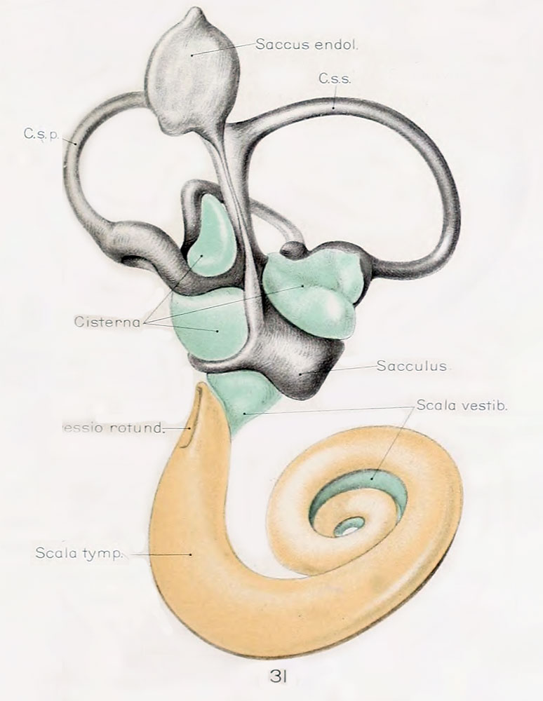
Fig. 31. Median view of left membranous labyrinth and the periotic spaces in a human fetus 130 mm CRL
| Median view of a wax-plate reconstruction of the left membranous labyrinth and the periotic spaces in a human fetus 130 mm. crown-rump length (Carnegie Collection, No. 1018), enlarged 11.4 diameters.
The cistern and scala vestibuli are shown in green and the scala tympani is shown in orange, as in the previous figures. The oval impression on the proximal end of the scala tympani corresponds to the fenestra cochleae (rotunda). As yet there is no conmiunication at this point between the scala tympani and subarachnoid spaces, such as is found in the adult and known as the aquaductus cochleae. The spaces making up the cistern cover almost the whole of the utricle and saccule except the places at which the nerves enter and a small part of the medial surface near the attachment of the appendage.
|
Abbreviations
|
Reference
Streeter G.L. The histogenesis and growth of the otic capsule and its contained periotic tissue-spaces in the human embryo Contributions to Embryology Carnegie Institution No.20 (1918) pp5-54, 4 text-figures and 4 plates.
File history
Click on a date/time to view the file as it appeared at that time.
| Date/Time | Thumbnail | Dimensions | User | Comment | |
|---|---|---|---|---|---|
| current | 21:53, 22 April 2012 | | 774 × 1,000 (79 KB) | Z8600021 (talk | contribs) | ==Fig. 31== Median view of same model shown in figure 30, enlarged 11.4 diameters. The oval impression on the proximal end of the scala tympani corresponds to the fenestra cochleae (rotunda). As yet there is no conmiunication at this point between the s |
You cannot overwrite this file.
File usage
The following 3 pages use this file: