File:Stage5 bf05.jpg
Original file (1,200 × 692 pixels, file size: 256 KB, MIME type: image/jpeg)
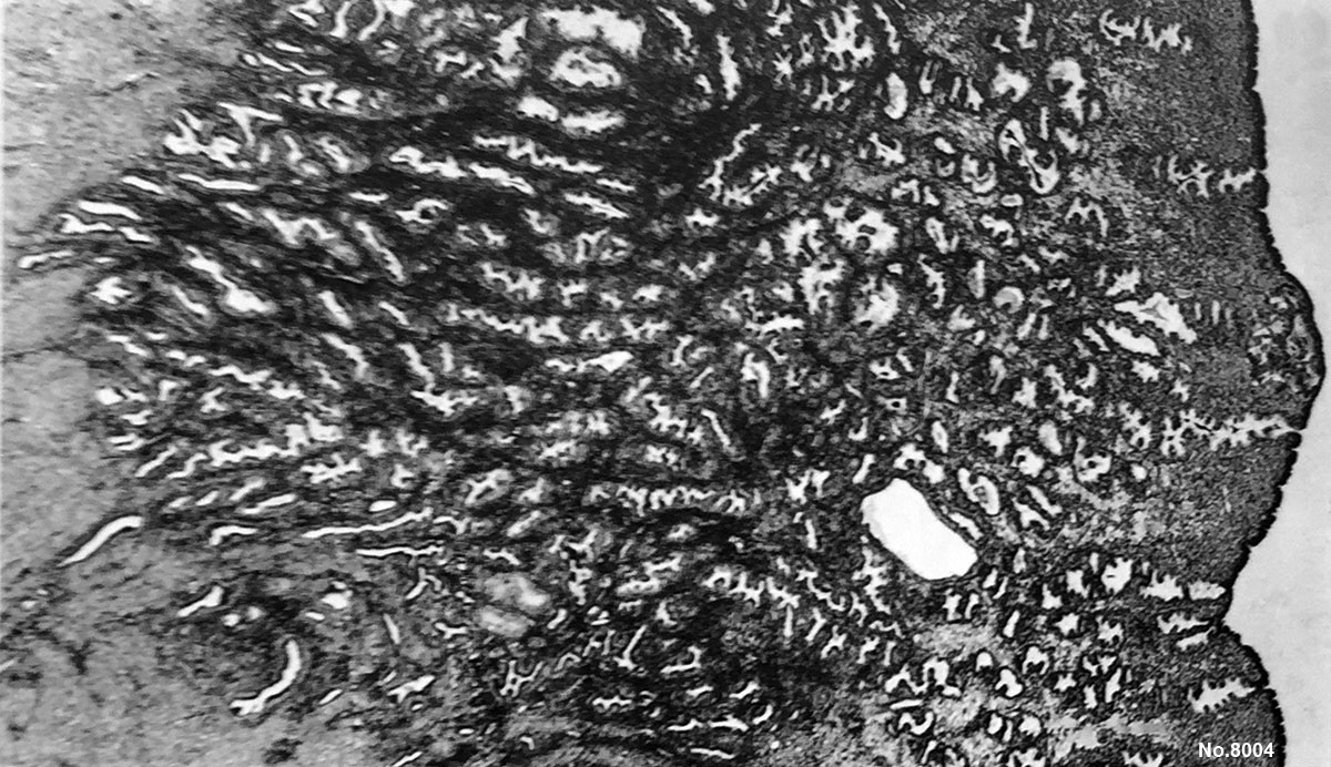
Human Embryo Carnegie Stage 5
Carnegie Collection Embryo No.8004 (stage 5b)
This histology section through the wall of the uterus shows the fully implanted conceptus located within the endometrium (far right, centre of image).
Under this the expanded uterine glands occupy most of the image. On the left of the image is the underlying myometrium.
- Links: Carnegie stage 5
Facts: Week 1-2, size 0.1 - 0.2 mm
Features: implantation completed, inner cell mass, bilaminar embryo, trophoblast development
- Hysterectomy
- Posterior wall of uterus
- Chorion - 0.582 x 0.45 mm
- Chorionic cavity - 0.312 x 0.185 mm
- Embryonic disc - 0.132 x 0.1 mm
- Stage 5 Links: Embryo No.8004 - Surface view | Embryo No.8004 - near implantation site | Embryo No.8004 - Stage 5b | Embryo No.8004 - Stage 5b labeled | Embryo No.8004 - Rotated view | Embryo No.7700 - implantation site | Embryo No.7700 - Stage 5c | Implantation | Week 2
- Carnegie Stages: 1 | 2 | 3 | 4 | 5 | 6 | 7 | 8 | 9 | 10 | 11 | 12 | 13 | 14 | 15 | 16 | 17 | 18 | 19 | 20 | 21 | 22 | 23 | About Stages | Timeline
Cite this page: Hill, M.A. (2024, April 24) Embryology Stage5 bf05.jpg. Retrieved from https://embryology.med.unsw.edu.au/embryology/index.php/File:Stage5_bf05.jpg
- © Dr Mark Hill 2024, UNSW Embryology ISBN: 978 0 7334 2609 4 - UNSW CRICOS Provider Code No. 00098G
File history
Click on a date/time to view the file as it appeared at that time.
| Date/Time | Thumbnail | Dimensions | User | Comment | |
|---|---|---|---|---|---|
| current | 17:51, 4 April 2015 | | 1,200 × 692 (256 KB) | Z8600021 (talk | contribs) | |
| 18:00, 23 September 2011 | 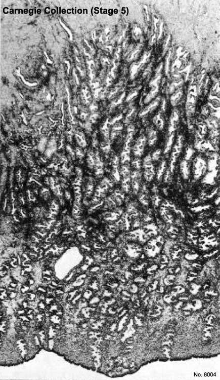 | 435 × 750 (122 KB) | S8600021 (talk | contribs) |
You cannot overwrite this file.
File usage
The following 5 pages use this file: