File:Stage13 otocyst.jpg

Original file (1,000 × 655 pixels, file size: 55 KB, MIME type: image/jpeg)
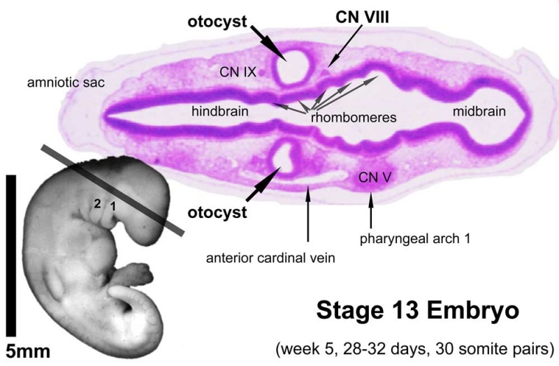
Week 5 - Otocyst
Stage 13 embryo (week 5) showing otocyst that will form the inner ear.
left Ventrolateral view of the whole embryo with 5-mm scale bar. At this stage of development no middle or external ear structures are apparent and will be derived later from pharyngeal arches one and two (labeled). The gray bar through the head indicates the plane of cross-section (right).
right A cross-section of the head showing the size and position of the otic vesicles. At this stage of development they lie within the head mesenchyme behind pharyngeal arch one and two and in close apposition to the developing hindbrain. Note the close position of the otic vesicle to the rhombomeres, hindbrain folds that represent the initial segmentation of the hindbrain. Also shown are developing cranial ganglia and blood vessel lying adjacent to the otic vesicles. The wall of the otic vesicle at this stage is a simple epithelium.
- Links: Inner Ear Development | Carnegie stage 13
Image Source: UNSW Embryology
Cite this page: Hill, M.A. (2024, April 19) Embryology Stage13 otocyst.jpg. Retrieved from https://embryology.med.unsw.edu.au/embryology/index.php/File:Stage13_otocyst.jpg
- © Dr Mark Hill 2024, UNSW Embryology ISBN: 978 0 7334 2609 4 - UNSW CRICOS Provider Code No. 00098G
File history
Click on a date/time to view the file as it appeared at that time.
| Date/Time | Thumbnail | Dimensions | User | Comment | |
|---|---|---|---|---|---|
| current | 00:10, 28 September 2009 | | 1,000 × 655 (55 KB) | S8600021 (talk | contribs) |
You cannot overwrite this file.
File usage
The following 18 pages use this file:
- 2009 Lecture 17
- 2010 BGD Practical 6 - Week 4
- 2010 Lab 3
- 2010 Lecture 17
- 2011 Lab 3 - Week 4
- AACP Meeting 2013 - Face Embryology
- ANAT2341 Lab 3 - Week 4
- ANAT2341 Lab 6 - Early Embryo
- BGDA Practical 7 - Week 4
- BGDB Face and Ear - Early Embryo
- BGD Lecture - Face and Ear Development
- Carnegie stage 13
- Hearing - Inner Ear Development
- Lecture - Sensory Development
- Neural - Cranial Nerve Development
- Sensory - Balance Development
- Sensory - Hearing and Balance Development
- Talk:2011 Lab 3