File:Stage10 sem6.jpg
Original file (614 × 1,000 pixels, file size: 57 KB, MIME type: image/jpeg)
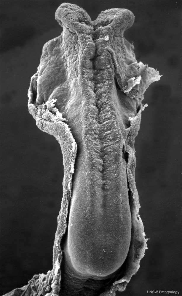
Human Embryo (Carnegie Stage 10)
Scanning electron micrograph dorsal view of Carnegie Stage 10 human embryo. This dorsal view of the early embryo mainly shows the neural groove before its closing to form the neural tube, the precursor of the spinal cord and brain.
- Week 4, 22 - 23 days, 2 - 3.5 mm, Somite number 4 - 12
- Neural groove forming from neural plate on upper surface.
- large brain fold region to top of image.
- narrow spinal cord region to bottom of image.
- Heart bulge can be seen on right ventral surface.
- Connecting stalk to the bottom of the image.
- Amniotic membrane cut edge shown at edge of developing embryo.
- Carnegie Stage 10 Links: vertical view | horizontal view | annotated | Carnegie stage 10 | Lecture - Early Neural | Neural System Development | Week 4
Image Source: Scanning electron micrographs of the Carnegie stages of the early human embryos are reproduced with the permission of Prof Kathy Sulik, from embryos collected by Dr. Vekemans and Tania Attié-Bitach. Images are for educational purposes only and cannot be reproduced electronically or in writing without permission.
- Carnegie Stages: 1 | 2 | 3 | 4 | 5 | 6 | 7 | 8 | 9 | 10 | 11 | 12 | 13 | 14 | 15 | 16 | 17 | 18 | 19 | 20 | 21 | 22 | 23 | About Stages | Timeline
Cite this page: Hill, M.A. (2024, April 20) Embryology Stage10 sem6.jpg. Retrieved from https://embryology.med.unsw.edu.au/embryology/index.php/File:Stage10_sem6.jpg
- © Dr Mark Hill 2024, UNSW Embryology ISBN: 978 0 7334 2609 4 - UNSW CRICOS Provider Code No. 00098G
File history
Click on a date/time to view the file as it appeared at that time.
| Date/Time | Thumbnail | Dimensions | User | Comment | |
|---|---|---|---|---|---|
| current | 12:32, 23 August 2009 | | 614 × 1,000 (57 KB) | S8600021 (talk | contribs) | Stage10day21somite4-5dorsalsem6v2.jpg |
You cannot overwrite this file.
File usage
The following 30 pages use this file:
- 2011 Lab 3 - Week 4
- ANAT2341 Lab 3 - Week 4
- ANAT3411 Neuroanatomy
- Brain Awareness Week 2012
- Carnegie stage 10
- Carnegie stage 10 gallery
- Cloaca Development
- Developmental Mechanism - Epithelial Invagination
- Human Embryo - Scanning electron microscopy
- Human Embryo SEM
- K12 Brain Awareness Week
- Lecture - Ectoderm Development
- Neural - Amygdala Development
- Neural - Basal Ganglia Development
- Neural - Cerebellum Development
- Neural - Diencephalon Development
- Neural - Medulla Oblongata Development
- Neural - Mesencephalon Development
- Neural - Metencephalon Development
- Neural - Myelencephalon Development
- Neural - Pons Development
- Neural - Prosencephalon Development
- Neural - Rhinencephalon Development
- Neural - Rhombencephalon Development
- Neural - Spinal Cord Development
- Neural - Tectum Development
- Neural - Telencephalon Development
- Neural - Thalamus Development
- Neural System Development
- Scanning Electron Microscopy