File:Spleen structure 02.jpg
From Embryology
No higher resolution available.
Spleen_structure_02.jpg (401 × 463 pixels, file size: 46 KB, MIME type: image/jpeg)
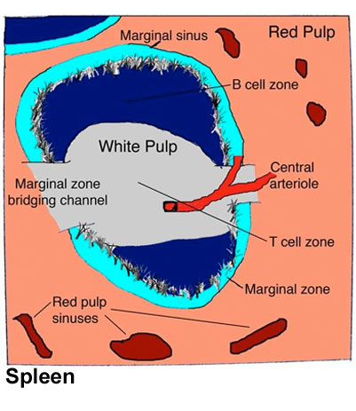
Spleen Structure Cartoon
- White pulp - consists of T cell zones (also known as the periarteriolar lymphoid sheath (PALS)) containing networks of fibroblastic reticular cells (FRC) surrounding a central arteriole, together with B cell follicles containing a central network of follicular dendritic cells (FDC).
- Immune cells enter the white pulp at regions where the T cell zones abut the MZ, known as the MZ bridging channels.
- Marginal zones - (MZ) surrounding the white pulp contain marginal reticular cells (MRC), particularly at the edges of the B cell follicles.
- Central arteriole - Blood and leukocytes entering the spleen pass through branches of the central arteriole, which end in the marginal sinuses and red pulp.
- Red pulp - In the cords of the red pulp, a dense network of reticular fibroblasts and fibres construct an open blood network, which is marked by its lack of a typical endothelial cell lining.
- Large numbers of macrophages phagocytose dying or damaged red blood cells in the red pulp (not shown).
Reference
<pubmed>19644499</pubmed>| PMC2785037 | Nat Rev Immunol.
File history
Click on a date/time to view the file as it appeared at that time.
| Date/Time | Thumbnail | Dimensions | User | Comment | |
|---|---|---|---|---|---|
| current | 18:55, 22 February 2012 | | 401 × 463 (46 KB) | Z8600021 (talk | contribs) |
You cannot overwrite this file.
File usage
The following 4 pages use this file: