File:Somite cartoon2.png
From Embryology
No higher resolution available.
Somite_cartoon2.png (400 × 300 pixels, file size: 16 KB, MIME type: image/png)
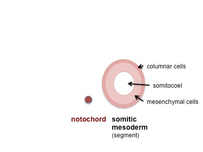
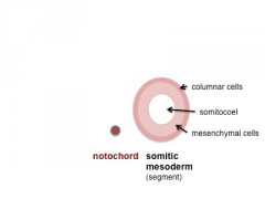
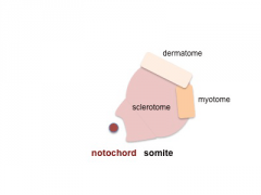
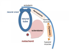
2. Somite Development - Early Somite
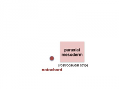
- Paraxial mesoderm towards the rostral (cranial) end, begins to segment forming the first somite.
- Somites are then sequentially added caudally in pairs.
- The somite surface cells undergo a mesenchymal to epithelial transition, forming a transient epithelial covering on each somite.
- The mesoderm under the surface remains as mesenchyme and proliferates.
- The early forming somites have a cavity (somitocoel) in their core that is lost as the somite matures.
Note - the cartoons show just the embryo righthand side mesoderm development (the same events occur on the lefthand side).
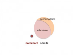
- Somite Links: 1 paraxial | 2 early somite | 3 sclerotome and dermomyotome | 4 dermatome and myotome | 5 somite spreading | SEM image - Human Embryo (week 4) showing somites | Movie - somitogenesis Hes expression
- Somite Cartoons
paraxial
early somite
sclerotome and dermomyotome
dermatome and myotome
somite spreading


Cite this page: Hill, M.A. (2024, April 20) Embryology Somite cartoon2.png. Retrieved from https://embryology.med.unsw.edu.au/embryology/index.php/File:Somite_cartoon2.png
- © Dr Mark Hill 2024, UNSW Embryology ISBN: 978 0 7334 2609 4 - UNSW CRICOS Provider Code No. 00098G
File history
Click on a date/time to view the file as it appeared at that time.
| Date/Time | Thumbnail | Dimensions | User | Comment | |
|---|---|---|---|---|---|
| current | 17:59, 16 May 2014 | | 400 × 300 (16 KB) | Z8600021 (talk | contribs) | |
| 10:41, 10 August 2009 | 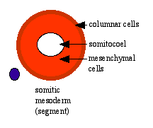 | 216 × 181 (3 KB) | MarkHill (talk | contribs) | Somite Development cartoon 2 Paraxial mesoderm towards the rostral end, begins to segment forming the first somite. Somites are then sequentially added caudally. The somitocoel, is a cavity forming in early somites, which is lost as the somite matures. |
You cannot overwrite this file.
File usage
The following 30 pages use this file:
- 2009 Lecture 5
- 2010 BGD Lecture - Development of the Embryo/Fetus 1
- 2010 BGD Lecture - Development of the Embryo/Fetus 2
- 2010 BGD Practical 6 - Week 3
- 2010 Lab 3
- 2010 Lecture 5
- 2011 Lab 3 - Week 3
- ANAT2341 Lab 3 - Week 3
- BGDA Lecture - Development of the Embryo/Fetus 1
- BGDA Lecture - Development of the Embryo/Fetus 2
- BGDA Practical 7 - Week 3
- Developmental Mechanism - Epithelial Mesenchymal Transition
- Lecture - Mesoderm Development
- Musculoskeletal System - Limb Development
- Musculoskeletal System Development
- Somite Musculoskeletal Movie
- Somitogenesis
- Talk:2010 BGD Practical 6 - Week 3
- Talk:2011 Lab 3
- File:Mesoderm cartoon 05.jpg
- File:Mesoderm cartoon 06.jpg
- File:Mesoderm cartoon 07.jpg
- File:Mesoderm cartoon 08.jpg
- File:Mesoderm cartoon 09.jpg
- File:Somite cartoon1.png
- File:Somite cartoon2.png
- File:Somite cartoon3.png
- File:Somite cartoon4.png
- File:Somite cartoon5.png
- Template:Somite cartoon