File:Skull CT normal sutures.jpg

Original file (1,000 × 900 pixels, file size: 138 KB, MIME type: image/jpeg)
Skull Normal Sutures

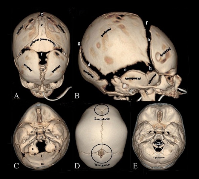
Computed Tomography (CT) scan with 3D surface-rendered reconstructions of different views of the newborn skull.
- Skull CT Images: Normal overview | Normal vertex and lateral | Normal endocranial and vertex | Normal Vertex - Fontanels | Dolichocephaly and Scaphocephaly | Coronal Synostosis | Anterior Plagiocephaly | Turricephaly | Posterior Plagiocephaly | Deformational Plagiocepahly | Trigonocephaly | Oxycephaly | Computed Tomography
A - Vertex view
B - Lateral view
- (a) Metopic suture; (b) coronal sutures; (c) sagittal suture; (d) lambdoid suture; (e) squamosal suture; (f) anterior fontanel; (g) posterior fontanel; (h) sphenoidal fontanel; (i) mastoid fontanel.
- Cranial vault bones usually ossify from the center to periphery, which results in this “widened” appearance of the sutures in the newborn.
C - Endocranial skull base view
Shows portions of the occipital bone and sutures
- (j) Basioccipital; (k) paired exoccipital; (l) supraoccipital; and (m) interparietal. Associated synchondroses are (n) spheno-occipital; (o)anterior intra-occipital; (p) posterior intra-occipital; (q) petro-occipital; (r) occipitomastoid; (s) and mendosal sutures. Note that o, k, p and s are paired structures.
D - Vertex view
Shows the lambda (point of intersection of the sagittal and lambdoid sutures) and bregma (point of intersection of the coronal and sagittal sutures.
E - Endocranial skull base view
Shows the basion (located on the basiocciput, at the midpoint of the anterior margin of the foramen magnum) and opisthion (located on the occipital bone, at the midpoint of the posterior margin of the foramen magnum).
- Skull Links: Skull Development | Historic - skull of a human fetus of 43 millimeters greatest length | Computed Tomography
Reference
Khanna PC, Thapa MM, Iyer RS & Prasad SS. (2011). Pictorial essay: The many faces of craniosynostosis. Indian J Radiol Imaging , 21, 49-56. PMID: 21431034 DOI.
Copyright
Paritosh C Khanna © 2007 - 2012 Indian Journal of Radiology and Imaging
This is an open-access article distributed under the terms of the Creative Commons Attribution License, which permits unrestricted use, distribution, and reproduction in any medium, provided the original work is properly cited. Attribution-NonCommercial-ShareAlike 3.0 Unported (CC BY-NC-SA 3.0)
Original file name: Figure 1(A-E): IJRI-21-49-g001.jpg http://www.ijri.org/viewimage.asp?img=IndianJRadiolImaging_2011_21_1_49_76055_f2.jpg resized and relabelled.
Cite this page: Hill, M.A. (2024, April 24) Embryology Skull CT normal sutures.jpg. Retrieved from https://embryology.med.unsw.edu.au/embryology/index.php/File:Skull_CT_normal_sutures.jpg
- © Dr Mark Hill 2024, UNSW Embryology ISBN: 978 0 7334 2609 4 - UNSW CRICOS Provider Code No. 00098G
File history
Click on a date/time to view the file as it appeared at that time.
| Date/Time | Thumbnail | Dimensions | User | Comment | |
|---|---|---|---|---|---|
| current | 08:01, 17 March 2012 | | 1,000 × 900 (138 KB) | Z8600021 (talk | contribs) | |
| 18:33, 23 May 2011 | 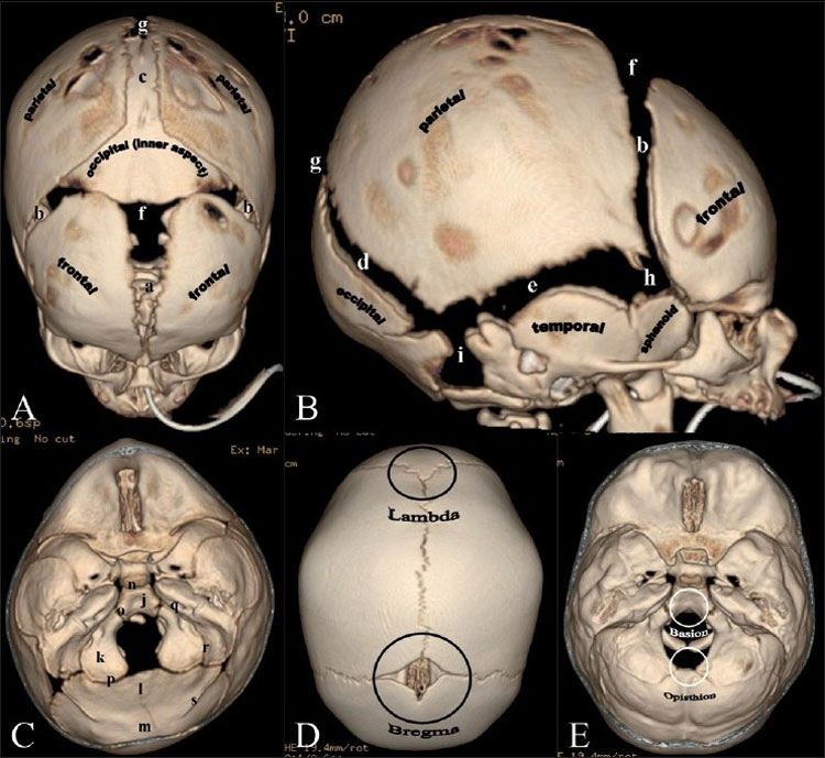 | 750 × 689 (105 KB) | S8600021 (talk | contribs) | ==Skull Normal Sutures== Computed Tomography (CT) scan with 3D surface-rendered reconstructions. Vertex (A) and lateral (B) views. (a) Metopic suture; (b) coronal sutures; (c) sagittal suture; (d) lambdoid suture; (e) squamosal suture; (f) anterior fonta |
You cannot overwrite this file.
File usage
The following 10 pages use this file:
- 2011 Lab 6 - Postnatal
- AACP Meeting 2013 - Face Embryology
- ANAT2341 Lab 6 - Postnatal
- BGDB Face and Ear - Postnatal
- BGD Lecture - Face and Ear Development
- Computed Tomography
- Head Development
- Lecture - Head Development
- Musculoskeletal System - Skull Development
- Neural Exam - Newborn head shape and sutures