File:Skeletal Muscle EM02.jpg
From Embryology
Size of this preview: 800 × 581 pixels. Other resolution: 2,191 × 1,590 pixels.
Original file (2,191 × 1,590 pixels, file size: 941 KB, MIME type: image/jpeg)
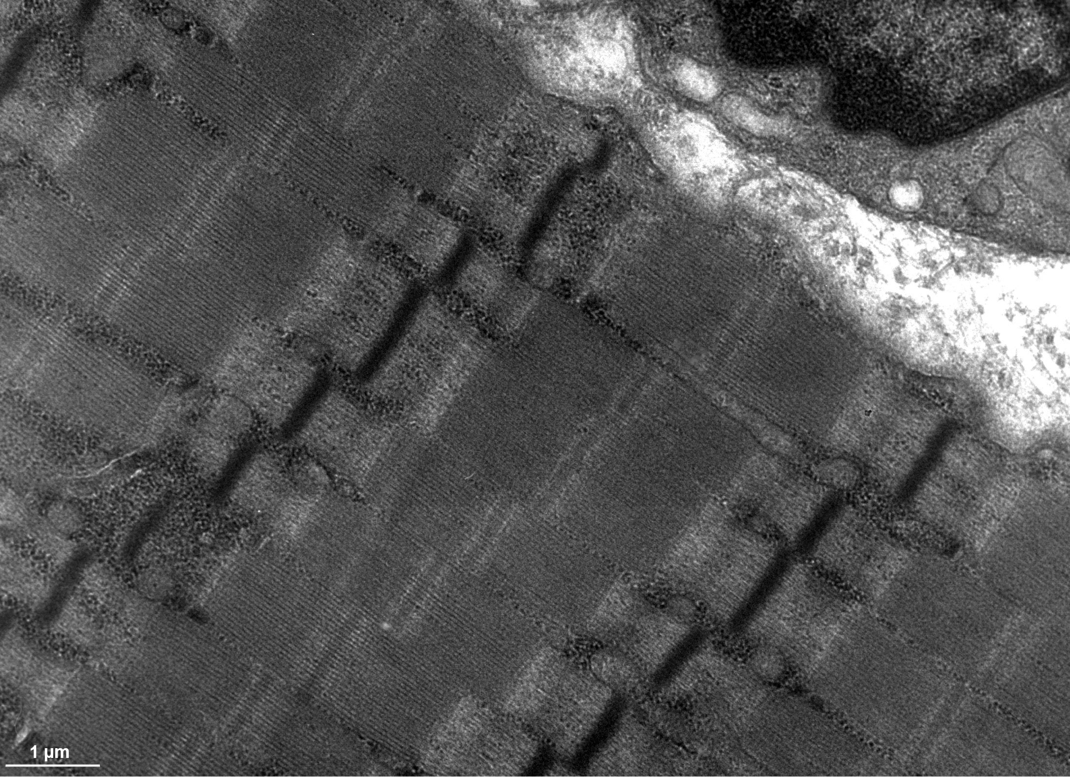
Skeletal Muscle EM
Transmission electron microscope image of a thin longitudinal section cut through an area of human skeletal muscle tissue. Image shows several myofibrils, each with the distinct banding pattern of individual sarcomeres.
- Skeletal Muscle EM Links: EM 1 | EM 2 | EM 3 | EM 4 | EM 5 | Virtual Slide 1 | Virtual Slide 2 | Virtual Slide 3 | Virtual Slide 4 | Virtual Slide 5 | Skeletal Muscle Development | Electron Microscopy Virtual Slides
Image Source: Contributed by Dartmouth College Electron Microscope Facility special thanks to Chuck Daghlian and Louisa Howard. Gallery. Original images may have been altered in size contrast and labelling. (These images are in the public domain)
File history
Click on a date/time to view the file as it appeared at that time.
| Date/Time | Thumbnail | Dimensions | User | Comment | |
|---|---|---|---|---|---|
| current | 16:40, 16 April 2014 | | 2,191 × 1,590 (941 KB) | Z8600021 (talk | contribs) | ==Skeletal Muscle EM== {{SkeletalMuscleEM links}} |
You cannot overwrite this file.
File usage
There are no pages that use this file.