File:Sheep oocyte 03.jpg
Original file (650 × 651 pixels, file size: 94 KB, MIME type: image/jpeg)
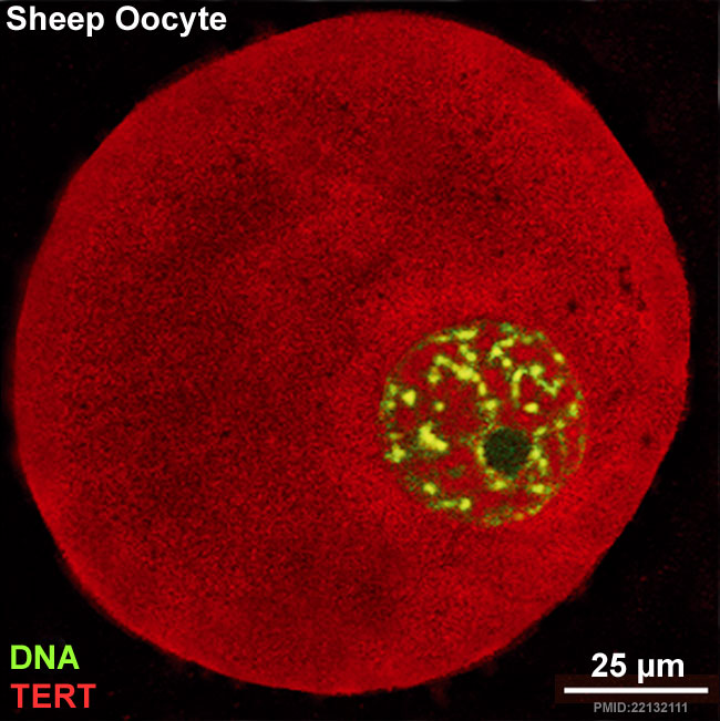
Sheep Oocyte Distribution of TERT
In vitro grown oocyte derived from an early antral follicle with a surrounded nucleolus (SN) chromatin configuration which reveals TERT between the nucleus and ooplasm.
- TERT - Red (Cy3-conjugated secondary antibody) (telomerase reverse transcriptase, TERT)
- DNA - Green (SYBR Green 14/I)
- Sheep Oocyte TERT: preantral | early antral | early antral | preovulatory follicle | Oocyte Development | Sheep Development
Reference
<pubmed>22132111</pubmed>| PLoS One.
Copyright: © 2011 Barboni et al. This is an open-access article distributed under the terms of the Creative Commons Attribution License, which permits unrestricted use, distribution, and reproduction in any medium, provided the original author and source are credited.
Figure 7. doi:info:doi/10.1371/journal.pone.0027550.g007
journal.pone.0027550.g007.jpg
Cropped, resized and relabelled from full figure.
Examples of oocyte TERT distribution, visualized with Cy3-conjugated secondary antibody (red), while DNA was counterstained in green with SYBR Green 14/I. TERT sub-cellular localization was influenced by chromatin configuration in all in vitro and in vivo classes of germ cells analyzed. (A,B) Oocytes derived from a preantral (PA) follicle (A) and an in vitro grown early antral (EA) follicle (B) with a not surrounded nucleolus (NSN) chromatin configuration which localized TERT in the nucleus. (C) in vitro grown oocyte derived from an EA follicle with a surrounded nucleolus (SN) chromatin configuration which reveals TERT between the nucleus and ooplasm. (D) Oocyte of a preovulatory follicle with condensed chromatin localized surrounding the nucleolus and the nuclear envelope (SNE configuration) and with a clear subcortical and perinuclear cytoplasmatic TERT distribution. Scale bar = 25 µm.
File history
Click on a date/time to view the file as it appeared at that time.
| Date/Time | Thumbnail | Dimensions | User | Comment | |
|---|---|---|---|---|---|
| current | 02:21, 7 June 2012 | | 650 × 651 (94 KB) | Z8600021 (talk | contribs) | ==Sheep Oocyte Distribution of TERT== TERT - telomerase catalytic subunit. Sheep oocyte 02.jpg {{Sheep Oocyte TERT}} ===Reference=== <pubmed>22132111</pubmed>| [http://www.plosone.org/article/info%3Adoi%2F10.1371%2Fjournal.pone.0027550 PLoS One.] Co |
You cannot overwrite this file.
File usage
The following 4 pages use this file: