File:Sea urchin ectoderm patterning model 01.jpg
Sea_urchin_ectoderm_patterning_model_01.jpg (300 × 528 pixels, file size: 28 KB, MIME type: image/jpeg)
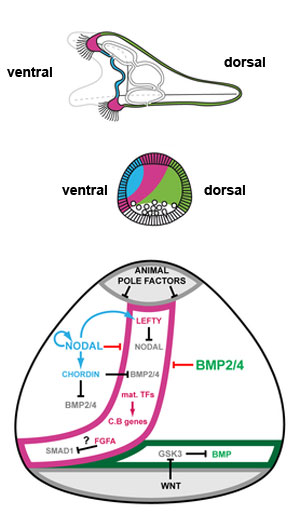
Changes in identity of ectodermal territories following perturbations of Nodal or BMP signaling and novel model of ectoderm patterning
Schemes describing the morphology of control embryos.
(A) control embryo. The thick ciliated epithelium of the ciliary band is restricted to a belt of cells at the interface between the ventral and dorsal ectoderm.
(F) Proposed model for regionalization of the ectoderm of the sea urchin embryo through restriction of the ciliary band fate by Nodal and BMP signaling. Maternal factors such as SoxB1 promote the early expression of ciliary band genes within the ectoderm. Nodal signaling on the ventral side promotes differentiation of the ventral ectoderm and stomodeum and represses the ciliary band fate probably through the activity of Goosecoid as well as of additional repressors. Nodal induces its antagonist Lefty, which diffuses away from the ventral ectoderm up to the presumptive ciliary band territory. Within the ventral ectoderm, Nodal induces expression of bmp2/4 and of its antagonist chordin. Chordin prevents BMP signaling within the ventral ectoderm and probably within the presumptive ciliary band region. At blastula stages, protein complexes containing BMP2/4 and Chordin can diffuse towards the dorsal side to specify dorsal fates. In the dorsal ectoderm, BMP signaling strongly repress the ciliary band fate partly by inducing the expression of the irxA repressor. A high level of MAP kinase activity resulting from FGFA signaling in the lateral ectoderm likely contributes to maintain a low level of Nodal and BMP signaling within the presumptive ciliary band region by phosphorylating Smad1/5/8 and Smad2/3 in the linker region, which inhibits their activity. The presence of Chordin and Lefty in the prospective ciliary band allows expression of ciliary band genes to be maintained in this region. The ectoderm surrounding the blastopore differentiates into dorsal ectoderm likely because it receives Wnt signals that antagonize GSK3 and promote BMP signaling. Original file name: Figure 11. Journal.pgen.1001259.g011.png
Reference
<pubmed>21203442</pubmed>
Citation: Saudemont A, Haillot E, Mekpoh F, Bessodes N, Quirin M, et al. (2010) Ancestral Regulatory Circuits Governing Ectoderm Patterning Downstream of Nodal and BMP2/4 Revealed by Gene Regulatory Network Analysis in an Echinoderm. PLoS Genet 6(12): e1001259. doi:10.1371/journal.pgen.1001259
Copyright: © 2010 Saudemont et al. This is an open-access article distributed under the terms of the Creative Commons Attribution License, which permits unrestricted use, distribution, and reproduction in any medium, provided the original author and source are credited.
File history
Click on a date/time to view the file as it appeared at that time.
| Date/Time | Thumbnail | Dimensions | User | Comment | |
|---|---|---|---|---|---|
| current | 17:56, 12 April 2011 | | 300 × 528 (28 KB) | S8600021 (talk | contribs) | ==Changes in identity of ectodermal territories following perturbations of Nodal or BMP signaling and novel model of ectoderm patterning== Schemes describing the morphology of control embryos. (A) control embryo. The thick ciliated epithelium of the ci |
You cannot overwrite this file.
File usage
The following page uses this file: