File:Sea urchin SEM03.jpg

Original file (1,000 × 712 pixels, file size: 154 KB, MIME type: image/jpeg)
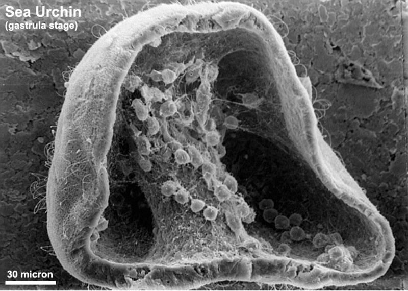
Sea Urchin Gastrula Stage
Scanning electron microscope image of the sea urchin (Strongylocentrotus drobachiensus) at the gastrula stage.
Embryo was cut in half to reveal blastocoel cavity, containing blastocoel matrix material, primary mesenchyme cells and secondary mesenchyme cells migrating from the tip of the archenteron.
Class Echinoidea - Superorder Echinacea - Order Echinoida - Strongylocentrotus drobachiensus
- Links: Sea Urchin Development
Image Source: Contributed by Dartmouth College Electron Microscope Facility special thanks to Chuck Daghlian and Louisa Howard. Gallery. Original images may have been altered in size contrast and labelling. (These images are in the public domain)
JEOL 35C SEM Evelyn Spiegel, Louisa Howard
File history
Click on a date/time to view the file as it appeared at that time.
| Date/Time | Thumbnail | Dimensions | User | Comment | |
|---|---|---|---|---|---|
| current | 14:13, 2 June 2011 | | 1,000 × 712 (154 KB) | S8600021 (talk | contribs) | ==Sea Urchin Gastrula Stage== Scanning electron microscope image of Strongylocentrotus drobachiensus (sea urchin) at the gastrula stage. Embryo was cut in half to reveal blastocoel cavity, containing blastocoel matrix material, primary mesenchyme cells |
You cannot overwrite this file.
File usage
The following page uses this file: