File:Rugh 018.jpg
From Embryology
Size of this preview: 800 × 566 pixels. Other resolution: 1,200 × 849 pixels.
Original file (1,200 × 849 pixels, file size: 192 KB, MIME type: image/jpeg)
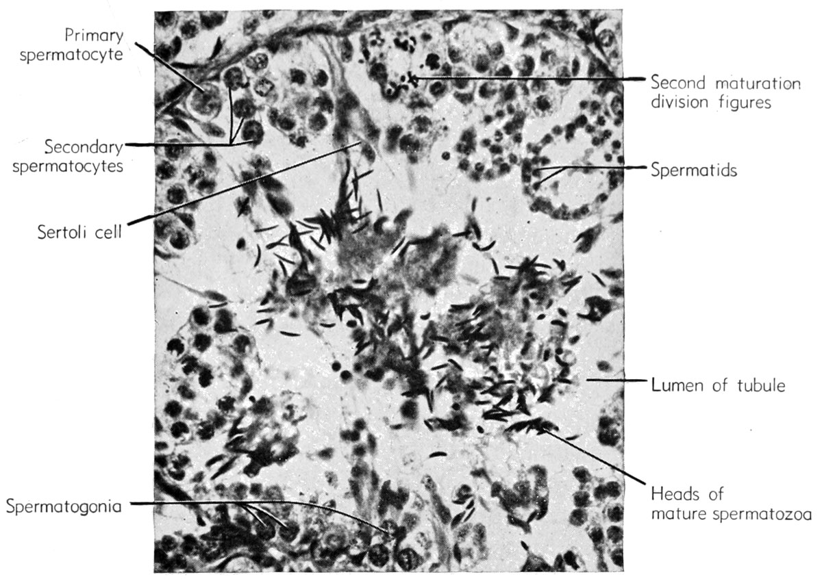
Spermatogenetic stages in the seminiterous tubule of the frog testis
| Historic Disclaimer - information about historic embryology pages |
|---|
 |
Reference
Rugh R. Book - The Frog Its Reproduction and Development. (1951) The Blakiston Company.
Cite this page: Hill, M.A. (2024, April 24) Embryology Rugh 018.jpg. Retrieved from https://embryology.med.unsw.edu.au/embryology/index.php/File:Rugh_018.jpg
- © Dr Mark Hill 2024, UNSW Embryology ISBN: 978 0 7334 2609 4 - UNSW CRICOS Provider Code No. 00098G
File history
Click on a date/time to view the file as it appeared at that time.
| Date/Time | Thumbnail | Dimensions | User | Comment | |
|---|---|---|---|---|---|
| current | 16:18, 6 April 2013 | | 1,200 × 849 (192 KB) | Z8600021 (talk | contribs) | Reverted to version as of 06:14, 6 April 2013 |
| 16:18, 6 April 2013 |  | 383 × 600 (15 KB) | Z8600021 (talk | contribs) | ||
| 16:14, 6 April 2013 |  | 1,200 × 849 (192 KB) | Z8600021 (talk | contribs) | ==Spermatogenetic stages in the seminiterous tubule of the frog testis== {{Rugh1951 footer}} |
You cannot overwrite this file.
File usage
The following 2 pages use this file: