File:Respiratory histology 12.jpg
Respiratory_histology_12.jpg (450 × 600 pixels, file size: 88 KB, MIME type: image/jpeg)
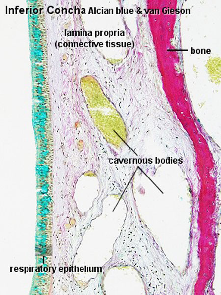
Inferior Concha Respiratory Histology
Inferior Concha, human
(Stain - Alcian blue)
(Stain - van Gieson)
Layers from surface epithelium to underlying bone.
Respiratory epithelium
- goblet cells
- ciliated cells
- basal cells
Lamina propria
- connective tissue
- cavernous sinusoids - large spaces (empty or filled with red blood cells)
- glandular tissue - mucous glands (green) and muco-serous glands (brownish-green)
Bone
- Lamellae and osteocytes in lacunae.
- Haversian systems are rare or absent.
- Respiratory Histology: Bronchiole | Alveolar Duct | Alveoli | EM Alveoli septum | Alveoli Elastin | Trachea 1 | Trachea 2 | labeled lung | unlabeled lung | Respiratory Bronchiole | Lung Reticular Fibres | Nasal Inferior Concha | Nasal Respiratory Epithelium | Olfactory Region overview | Olfactory Region Epithelium | Histology Stains
Links: Histology | Histology Stains | Blue Histology images copyright Lutz Slomianka 1998-2009. The literary and artistic works on the original Blue Histology website may be reproduced, adapted, published and distributed for non-commercial purposes. See also the page Histology Stains.
Cite this page: Hill, M.A. (2024, April 19) Embryology Respiratory histology 12.jpg. Retrieved from https://embryology.med.unsw.edu.au/embryology/index.php/File:Respiratory_histology_12.jpg
- © Dr Mark Hill 2024, UNSW Embryology ISBN: 978 0 7334 2609 4 - UNSW CRICOS Provider Code No. 00098G
File history
Click on a date/time to view the file as it appeared at that time.
| Date/Time | Thumbnail | Dimensions | User | Comment | |
|---|---|---|---|---|---|
| current | 23:03, 28 February 2012 | | 450 × 600 (88 KB) | Z8600021 (talk | contribs) |
You cannot overwrite this file.