File:Respiratory histology 02.jpg
From Embryology
No higher resolution available.
Respiratory_histology_02.jpg (450 × 600 pixels, file size: 37 KB, MIME type: image/jpeg)
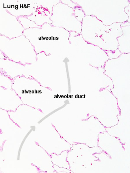
Respiratory Duct and Alveoli
- Respiratory bronchioles divide and terminate in alveolar ducts.
- alveolar duct "walls" consists of entirely of alveoli.
- Respiratory Histology: Bronchiole | Alveolar Duct | Alveoli | EM Alveoli septum | Alveoli Elastin | Trachea 1 | Trachea 2 | labeled lung | unlabeled lung | Respiratory Bronchiole | Lung Reticular Fibres | Nasal Inferior Concha | Nasal Respiratory Epithelium | Olfactory Region overview | Olfactory Region Epithelium | Histology Stains
Links: Histology | Histology Stains | Blue Histology images copyright Lutz Slomianka 1998-2009. The literary and artistic works on the original Blue Histology website may be reproduced, adapted, published and distributed for non-commercial purposes. See also the page Histology Stains.
Cite this page: Hill, M.A. (2024, April 20) Embryology Respiratory histology 02.jpg. Retrieved from https://embryology.med.unsw.edu.au/embryology/index.php/File:Respiratory_histology_02.jpg
- © Dr Mark Hill 2024, UNSW Embryology ISBN: 978 0 7334 2609 4 - UNSW CRICOS Provider Code No. 00098G
Respiratory histology 02.jpg
Original file name: Lun10he.jpg
File history
Click on a date/time to view the file as it appeared at that time.
| Date/Time | Thumbnail | Dimensions | User | Comment | |
|---|---|---|---|---|---|
| current | 19:43, 27 February 2012 | | 450 × 600 (37 KB) | Z8600021 (talk | contribs) | |
| 15:49, 2 March 2011 | 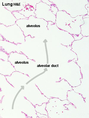 | 300 × 400 (24 KB) | S8600021 (talk | contribs) | ||
| 15:46, 2 March 2011 | 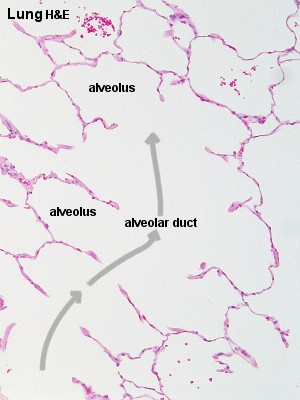 | 300 × 400 (30 KB) | S8600021 (talk | contribs) | ==Respiratory Bronchiole== Respiratory histology 02.jpg Original file name: Lun10he.jpg {{Template:Blue Histology}} Category:Respiratory |
You cannot overwrite this file.