File:Proboscis histology.jpg

Original file (600 × 1,041 pixels, file size: 166 KB, MIME type: image/jpeg)
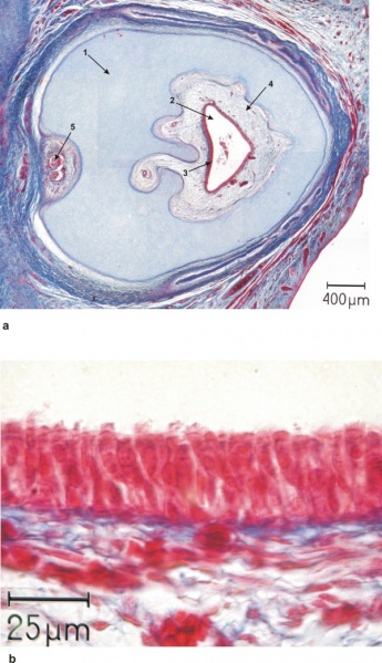
Holoprosencephaly - Histology of Proboscis
a) Overview of a histological section of the proboscis showing:
- closed circular cartilaginous wall
- with a central canal
- with an epithelial lining
- supported by loose connective tissue
- CT contains blood vessels and two nerves at the posterior side
b) Higher magnification of the epithelial lining of the central canal, depicting a respiratory epithelium with cilia (arrow 1) and goblet cells (arrow 2).
The head from a human fetus at 20 weeks gestation and a diagnosis of holoprosencephaly and cyclopia was investigated histologically and three-dimensionally reconstructed with CAD techniques. The cranial bones, blood vessels, nerves, eye and brain anlagen were reconstructed.
- "Holoprosencephaly seems to be a malformation of the most rostral parts of the brain and the cranium, as it affects mainly the telencephalon and the upper portion of the facial cranium. From the results of this investigation, and in combination with previous reports, it can be concluded that there is a defect in the definition of the median plane in embryonic development.
The histological finding of the proboscis with a respiratory epithelial lining leads to the conclusion that it is a nose-like structure and part of the fronto-nasal process which failed to develop properly. It further can be concluded that the ethmoid plays an important role in the development of the bilateral symmetry of the face, and may play an important role in the determination of the median plane."
International Classification of Diseases - Q04 Other congenital malformations of brain - Q04.2 Holoprosencephaly
- Neural Abnormality Links: Human holoprosencephaly cyclopia dissection | Proboscis histology | Holoprosencephaly | Neural Abnormalities | Sonic hedgehog
Reference
Arnold WH & Meiselbach V. (2009). 3-D reconstruction of a human fetus with combined holoprosencephaly and cyclopia. Head Face Med , 5, 14. PMID: 19563629 DOI.
Copyright
Copyright © 2009 Arnold and Meiselbach; licensee BioMed Central Ltd.
This is an Open Access article distributed under the terms of the Creative Commons Attribution License (http://creativecommons.org/licenses/by/2.0), which permits unrestricted use, distribution, and reproduction in any medium, provided the original work is properly cited.
Head Face Med. 2009; 5: 14. Published online 2009 June 29. doi: 10.1186/1746-160X-5-14.
Link: http://www.pubmedcentral.nih.gov/articlerender.fcgi?artid=2709107&rendertype=figure&id=F9
Original File name: 1746-160X-5-14-9
Cite this page: Hill, M.A. (2024, April 24) Embryology Proboscis histology.jpg. Retrieved from https://embryology.med.unsw.edu.au/embryology/index.php/File:Proboscis_histology.jpg
- © Dr Mark Hill 2024, UNSW Embryology ISBN: 978 0 7334 2609 4 - UNSW CRICOS Provider Code No. 00098G
File history
Click on a date/time to view the file as it appeared at that time.
| Date/Time | Thumbnail | Dimensions | User | Comment | |
|---|---|---|---|---|---|
| current | 08:47, 9 August 2009 | | 600 × 1,041 (166 KB) | S8600021 (talk | contribs) | Histology of proboscis. a) Overview of a histological section of the proboscis showing the closed circular cartilaginous wall (arrow 1) with a central canal (arrow 2) with an epithelial lining (arrow 3) supported by loose connective tissue (arrow 4) whi |
You cannot overwrite this file.
File usage
The following 2 pages use this file: