File:Plasma cell clockface nucleus 01.jpg
Plasma_cell_clockface_nucleus_01.jpg (400 × 400 pixels, file size: 27 KB, MIME type: image/jpeg)
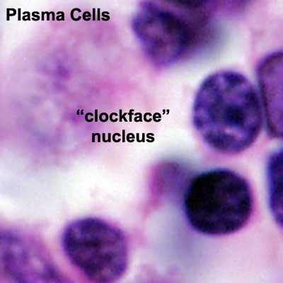
Plasma Cell "clockface" Nucleus
Nucleus has darker (heterochromatin) regions around periphery of nucleus separated by lighter (euchromatin) regions. This gives the stained nucleus the generic appearance of a "clock face" (not the correct scientific name).
- Heterochromatin - tightly packed form of DNA
- Euchromatin - loosely packed form of DNA.
Alternative nomenclature - activated B cell, plasma B cells, plasmocytes, effector B cells and B cell that is secreting antibody.
(Stain - Haematoxylin Eosin)
- Immune Images: Oesophagus MALT | Colon MALT | Peyer's patch overview | Peyer's patch detail | Cartoon - IEL development | Cartoon - IEL function | Cartoon - IEL differentiation | Mesenteric Lymph Nodes overview | Palatine Tonsil | Tonsil | Immune System Development
Links: Histology | Histology Stains | Blue Histology images copyright Lutz Slomianka 1998-2009. The literary and artistic works on the original Blue Histology website may be reproduced, adapted, published and distributed for non-commercial purposes. See also the page Histology Stains.
Cite this page: Hill, M.A. (2024, April 25) Embryology Plasma cell clockface nucleus 01.jpg. Retrieved from https://embryology.med.unsw.edu.au/embryology/index.php/File:Plasma_cell_clockface_nucleus_01.jpg
- © Dr Mark Hill 2024, UNSW Embryology ISBN: 978 0 7334 2609 4 - UNSW CRICOS Provider Code No. 00098G
File history
Click on a date/time to view the file as it appeared at that time.
| Date/Time | Thumbnail | Dimensions | User | Comment | |
|---|---|---|---|---|---|
| current | 14:22, 25 February 2013 | | 400 × 400 (27 KB) | Z8600021 (talk | contribs) | ==Plasma_cell_clockface_nucleus== {{Immune Images 2}} {{Blue Histology}} pey101he.jpg Category:Immune Category:Gastrointestinal Tract |
You cannot overwrite this file.
File usage
The following 3 pages use this file: