File:Placental villi 4.jpg
From Embryology

Size of this preview: 750 × 600 pixels. Other resolution: 1,280 × 1,024 pixels.
Original file (1,280 × 1,024 pixels, file size: 89 KB, MIME type: image/jpeg)
Human Term Placental Villi
- Placental villi at term.
- Surface layer of vill now lack cytotrophoblast cell layer and now are covered with only a syncitotrophoblast layer (shell).
- Syncitotrophoblast layer is variable in thickness from a thin cytoplasmic layer to regions of clumped nuclei.
- Vill core mainly occupied by fetal capillaries and larger blood vessels, with only a small amount of connective tissue.
- Fetal red blood cells now appear to lack the earlier distinctive nuclei.
- Villi Histology: First trimester (overview | villi | detail) | fetal and maternal RBCs | Term (overview | villi | detail) | Cord Histology | Placenta Histology | Placenta Development
Links: Histology | Histology Stains | Blue Histology images copyright Lutz Slomianka 1998-2009. The literary and artistic works on the original Blue Histology website may be reproduced, adapted, published and distributed for non-commercial purposes. See also the page Histology Stains.
Cite this page: Hill, M.A. (2024, April 23) Embryology Placental villi 4.jpg. Retrieved from https://embryology.med.unsw.edu.au/embryology/index.php/File:Placental_villi_4.jpg
- © Dr Mark Hill 2024, UNSW Embryology ISBN: 978 0 7334 2609 4 - UNSW CRICOS Provider Code No. 00098G
original file name Pll20he.jpg
File history
Click on a date/time to view the file as it appeared at that time.
| Date/Time | Thumbnail | Dimensions | User | Comment | |
|---|---|---|---|---|---|
| current | 02:42, 1 April 2012 | | 1,280 × 1,024 (89 KB) | Z8600021 (talk | contribs) | |
| 17:54, 30 March 2012 | 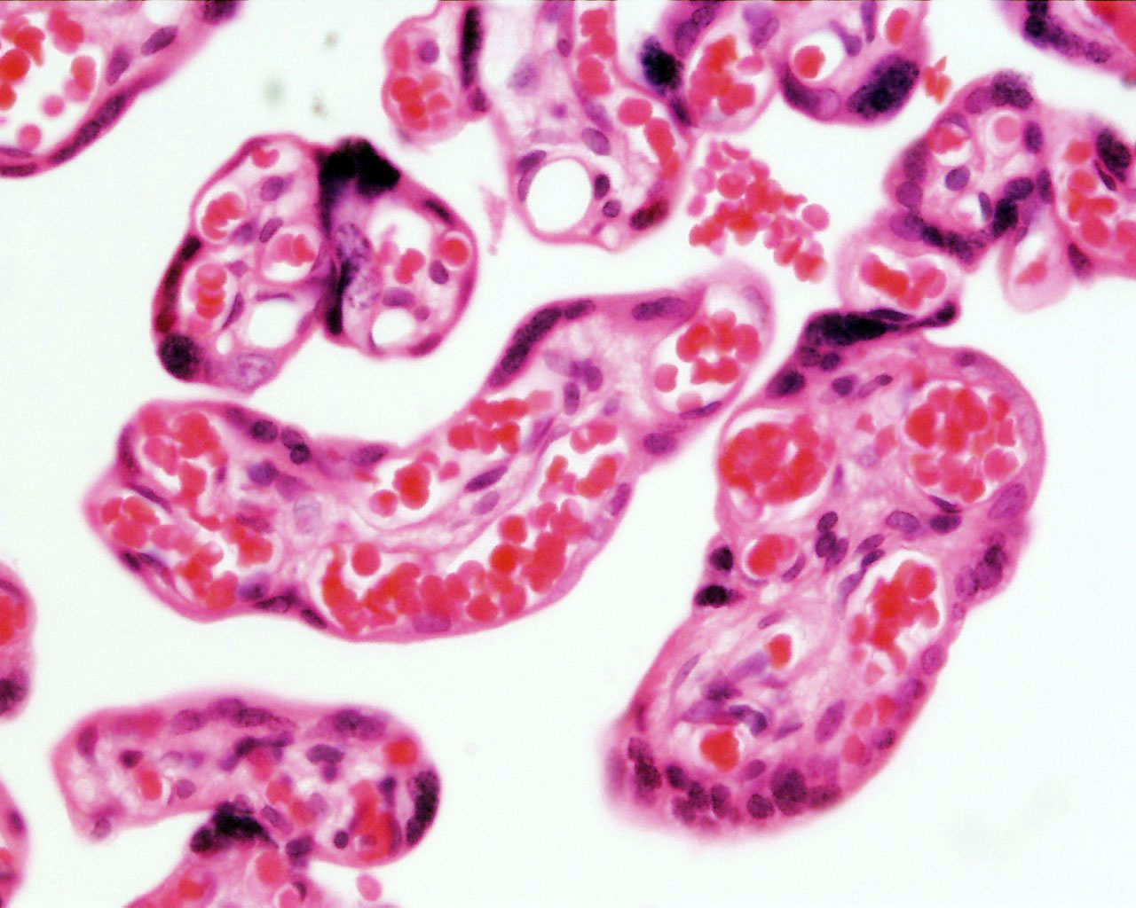 | 1,280 × 1,024 (206 KB) | Z8600021 (talk | contribs) | ||
| 16:38, 3 August 2009 | 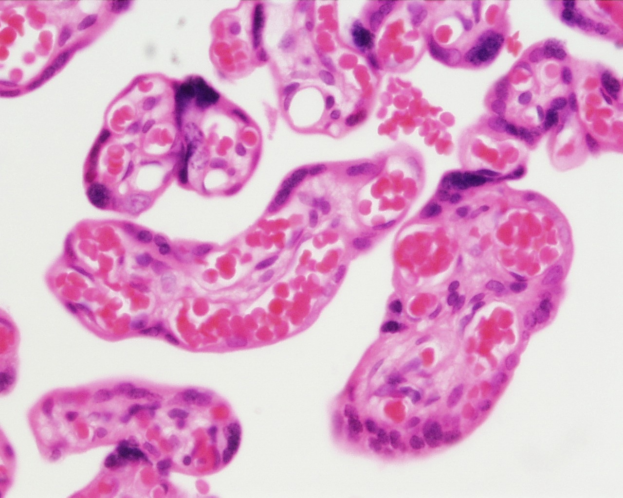 | 1,280 × 1,024 (226 KB) | MarkHill (talk | contribs) | Human placental villi cross-section placenta, term, human H&E x40 reproductive system, female, chorionic villi original file name Pll20he.jpg Image Source: UWA Blue Histology http://www.lab.anhb.uwa.edu.au/mb140/CorePages/FemaleRepro/femalerepro.htm |
You cannot overwrite this file.