File:Placental trophospongium.jpg
Placental_trophospongium.jpg (567 × 344 pixels, file size: 94 KB, MIME type: image/jpeg)
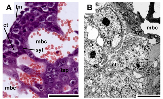
Rodent Placenta Histology
Placental trophospongium in initial pregnancy (Galea spixii) related to the guinea pig, has placental similarities to those of humans.
- A - HE The placenta mostly consists of trophospongium (tsp) around the maternal blood channels (mbc). Layers of cellular trophoblast (ct) are situated on fetal mesenchyme along the central excavation, covered by syncytial trophoblast (syt).
- B - TEM The trophoblast cells have large intercellular spaces in between and towards the syncytiotrophoblast.
Scale bars = 0.1 mm for histology and 2 μm for TEM.
- Links: Placenta - Histology
Reference
Oliveira MF, Mess A, Ambrósio CE, Dantas CA, Favaron PO & Miglino MA. (2008). Chorioallantoic placentation in Galea spixii (Rodentia, Caviomorpha, Caviidae). Reprod. Biol. Endocrinol. , 6, 39. PMID: 18771596 DOI.
Reprod Biol Endocrinol. 2008; 6: 39. Published online 2008 September 4. doi: 10.1186/1477-7827-6-39.
Copyright
© 2008 Oliveira et al; licensee BioMed Central Ltd.
This is an Open Access article distributed under the terms of the Creative Commons Attribution License (http://creativecommons.org/licenses/by/2.0), which permits unrestricted use, distribution, and reproduction in any medium, provided the original work is properly cited.
1477-7827-6-39-3.jpg
Cite this page: Hill, M.A. (2024, April 20) Embryology Placental trophospongium.jpg. Retrieved from https://embryology.med.unsw.edu.au/embryology/index.php/File:Placental_trophospongium.jpg
- © Dr Mark Hill 2024, UNSW Embryology ISBN: 978 0 7334 2609 4 - UNSW CRICOS Provider Code No. 00098G
File history
Click on a date/time to view the file as it appeared at that time.
| Date/Time | Thumbnail | Dimensions | User | Comment | |
|---|---|---|---|---|---|
| current | 09:25, 16 August 2009 | | 567 × 344 (94 KB) | S8600021 (talk | contribs) | Placental trophospongium in initial pregnancy. (A) HE. The placenta mostly consists of trophospongium (tsp) around the maternal blood channels (mbc). Layers of cellular trophoblast (ct) are situated on fetal mesenchyme along the central excavation, cove |
You cannot overwrite this file.
File usage
The following 4 pages use this file: