File:Pituitary histology 010.jpg
Original file (1,005 × 961 pixels, file size: 249 KB, MIME type: image/jpeg)
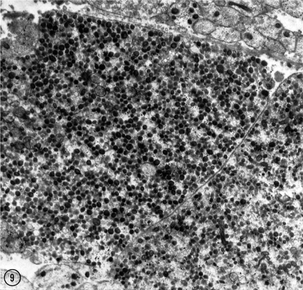
Pituitary Neurohypophysis Herring Bodies electron micrograph
The lobule of the opossum neurohypophysis is divided into three regions: a hilar, a palisade, and a septal zone.
This micrograph of the hilar region demonstrates two adjacent Herring bodies that occupy a major portion of the field. They are large bodies packed with neurosecretory granules that have been described as end bulb formations of axons.
At the upper right and lower left are fascicles of unmyelinated axons. Lead stained. X 13,500.
Reference
<pubmed>14128048 </pubmed>| PMC2106401 | J Cell Biol.
Copyright
Rockefeller University Press - Copyright Policy This article is distributed under the terms of an Attribution–Noncommercial–Share Alike–No Mirror Sites license for the first six months after the publication date (see http://www.jcb.org/misc/terms.shtml). After six months it is available under a Creative Commons License (Attribution–Noncommercial–Share Alike 4.0 Unported license, as described at https://creativecommons.org/licenses/by-nc-sa/4.0/ ). (More? Help:Copyright Tutorial)
File history
Click on a date/time to view the file as it appeared at that time.
| Date/Time | Thumbnail | Dimensions | User | Comment | |
|---|---|---|---|---|---|
| current | 23:46, 15 May 2012 | | 1,005 × 961 (249 KB) | Z8600021 (talk | contribs) | ==Pituitary Neurohypophysis Herring Bodies electron micrograph== |
You cannot overwrite this file.
File usage
The following page uses this file: