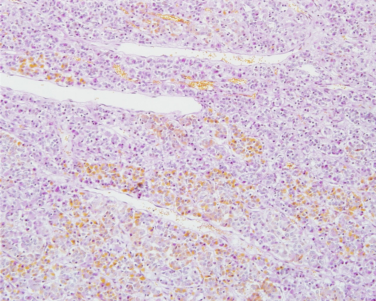
File:Pituitary histology 006.jpg
Original file (1,280 × 1,024 pixels, file size: 450 KB, MIME type: image/jpeg)
Pituitary Histology - Adenohypophysis
Acidophil cells
- about 65% of all cells.
- rounded and smaller than basophil cells (other stains identify subtypes).
- Somatotrophs - produce growth hormone (GH or somatotropin), stimulates liver cells to produce polypeptide growth factors which stimulate growth (stain with orange G)
- Mammotrophs - (lactotrophs) produce prolactin, maternal numbers increase in third trimester and postnatally in early lactation.
Basophil cells
Based on their hormone products basophils are divided into three subtypes (PAS stain all types reddish).
- Thyrotrophs - produce thyroid stimulating hormone (TSH or thyrotropin).
- Gonadotrophs - produce follicle stimulating hormone (FSH) and luteinizing hormone (LH)
- FSH stimulates in the male seminiferous tubule and female early follicular growth.
- LH stimulates male Leydig cell testosterone production and female oestrogen (estrogen) production, late follicular maturation, formation of corpus luteum.
- Corticotrophs - (or adrenocorticolipotrophs) produce adrenocorticotropic hormone (ACTH or corticotropin) and lipotropin (LPH).
- cell type in the pars intermedia where ACTH and LPH precursor undergoes hydrolysis into melanocyte stimulating hormone (MSH) and other peptides.
Chromophobe cells
- cells are unstained or weakly stained cells.
- either acidophils or basophils in a dormant or recently degranulated stage.
- may also include the secretory stem cells.
Pituitary, sheep - (Stain Periodic acid-Schiff / Orange G)
Identify acidophils, basophils and chromophobes and verify that the relative frequencies of the cells are different in different parts of the adenohypophysis.
- Pituitary Histology: Pituitary overview | Anterior H&E | Anterior H&E | Anterior labeled | PAS/O Overview | Acidophils | Basophils | Posterior labeled | Posterior unlabeled | Histology Stains | BGD - Endocrine Histology | Pituitary Development
Links: Histology | Histology Stains | Blue Histology images copyright Lutz Slomianka 1998-2009. The literary and artistic works on the original Blue Histology website may be reproduced, adapted, published and distributed for non-commercial purposes. See also the page Histology Stains.
Cite this page: Hill, M.A. (2024, April 23) Embryology Pituitary histology 006.jpg. Retrieved from https://embryology.med.unsw.edu.au/embryology/index.php/File:Pituitary_histology_006.jpg
- © Dr Mark Hill 2024, UNSW Embryology ISBN: 978 0 7334 2609 4 - UNSW CRICOS Provider Code No. 00098G
Original File Name: Hya10po.jpg
File history
Click on a date/time to view the file as it appeared at that time.
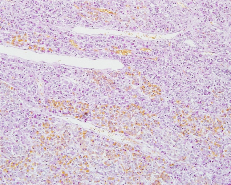
| Date/Time | Thumbnail | Dimensions | User | Comment | |
|---|---|---|---|---|---|
| current | 22:01, 15 May 2012 | | 1,280 × 1,024 (450 KB) | Z8600021 (talk | contribs) | ==Pituitary Histology - Adenohypophysis== ===Acidophil cells=== * about 65% of all cells. * rounded and smaller than basophil cells (other stains identify subtypes). * Somatotrophs - produce growth hormone (GH or somatotropin), stimulates liver cells to |
You cannot overwrite this file.
File usage
The following 2 pages use this file: