File:Patent ductus arteriosus angiogram.jpg
Original file (1,000 × 897 pixels, file size: 91 KB, MIME type: image/jpeg)
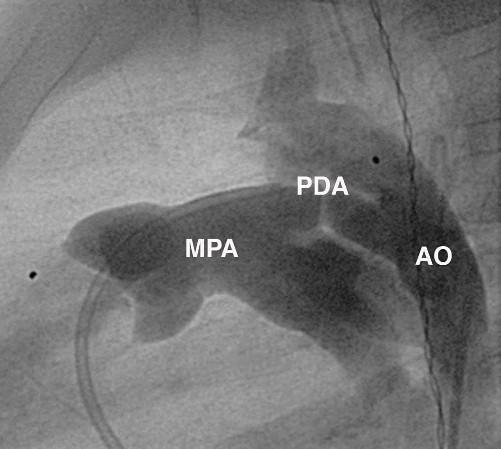
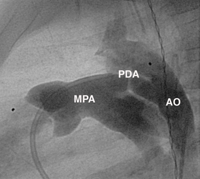
Patent Ductus Arteriosus (PDA) Angiogram
Lateral angiogram demonstrating a large tubular type duct (type C) with aortic (AO) to pulmonary flow (MPA).
The catheter can be seen entering the MPA from the right ventricle, a second pigtail catheter is positioned in the descending aorta from which contrast is delivered.
- Links: Cardiovascular System - Abnormalities | Patent Ductus Arteriosus cartoon | Patent ductus arteriosus classification | PDA echocardiogram | PDA angiogram
Reference
Forsey JT, Elmasry OA & Martin RP. (2009). Patent arterial duct. Orphanet J Rare Dis , 4, 17. PMID: 19591690 DOI.
Copyright
© 2009 Forsey et al; licensee BioMed Central Ltd. This is an Open Access article distributed under the terms of the Creative Commons Attribution License (http://creativecommons.org/licenses/by/2.0), which permits unrestricted use, distribution, and reproduction in any medium, provided the original work is properly cited. Original file name: 1750-1172-4-17-2.jpg http://www.ojrd.com/content/4/1/17/figure/F2
Cite this page: Hill, M.A. (2024, April 23) Embryology Patent ductus arteriosus angiogram.jpg. Retrieved from https://embryology.med.unsw.edu.au/embryology/index.php/File:Patent_ductus_arteriosus_angiogram.jpg
- © Dr Mark Hill 2024, UNSW Embryology ISBN: 978 0 7334 2609 4 - UNSW CRICOS Provider Code No. 00098G
File history
Click on a date/time to view the file as it appeared at that time.
| Date/Time | Thumbnail | Dimensions | User | Comment | |
|---|---|---|---|---|---|
| current | 12:44, 27 April 2011 | | 1,000 × 897 (91 KB) | S8600021 (talk | contribs) | ==Patent Ductus Arteriosus (PDA) Angiogram== ELateral angiogram demonstrating a large tubular type duct (type C) with aortic (AO) to pulmonary flow (MPA). The catheter can be seen entering the MPA from the right ventricle, a second pigtail catheter is po |
You cannot overwrite this file.