File:Parotid histology stratified columnar 01.jpg
From Embryology
No higher resolution available.
Parotid_histology_stratified_columnar_01.jpg (480 × 600 pixels, file size: 61 KB, MIME type: image/jpeg)
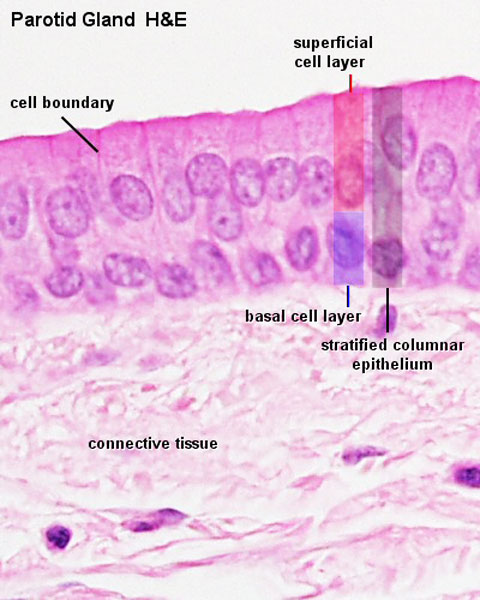
Parotid Gland Histology
Parotid gland epithelium is an example of a stratified columnar epithelium.
- lie outside the oral cavity
- tubuloacinar glands
- almost all serous acini
- Parotid Gland Links: Gland overview | lobe overview | serous acini | Striated duct and serous | Intercalated duct | excretory duct | Gland epithelium | Duct cartoon
File history
Click on a date/time to view the file as it appeared at that time.
| Date/Time | Thumbnail | Dimensions | User | Comment | |
|---|---|---|---|---|---|
| current | 13:01, 26 March 2012 | | 480 × 600 (61 KB) | Z8600021 (talk | contribs) | ==Parotid Gland Histology== Parotid gland epithelium is an example of a stratified columnar epithelium. |
You cannot overwrite this file.
File usage
The following 4 pages use this file: