File:Ovary histology 006.jpg
Original file (1,280 × 1,024 pixels, file size: 424 KB, MIME type: image/jpeg)
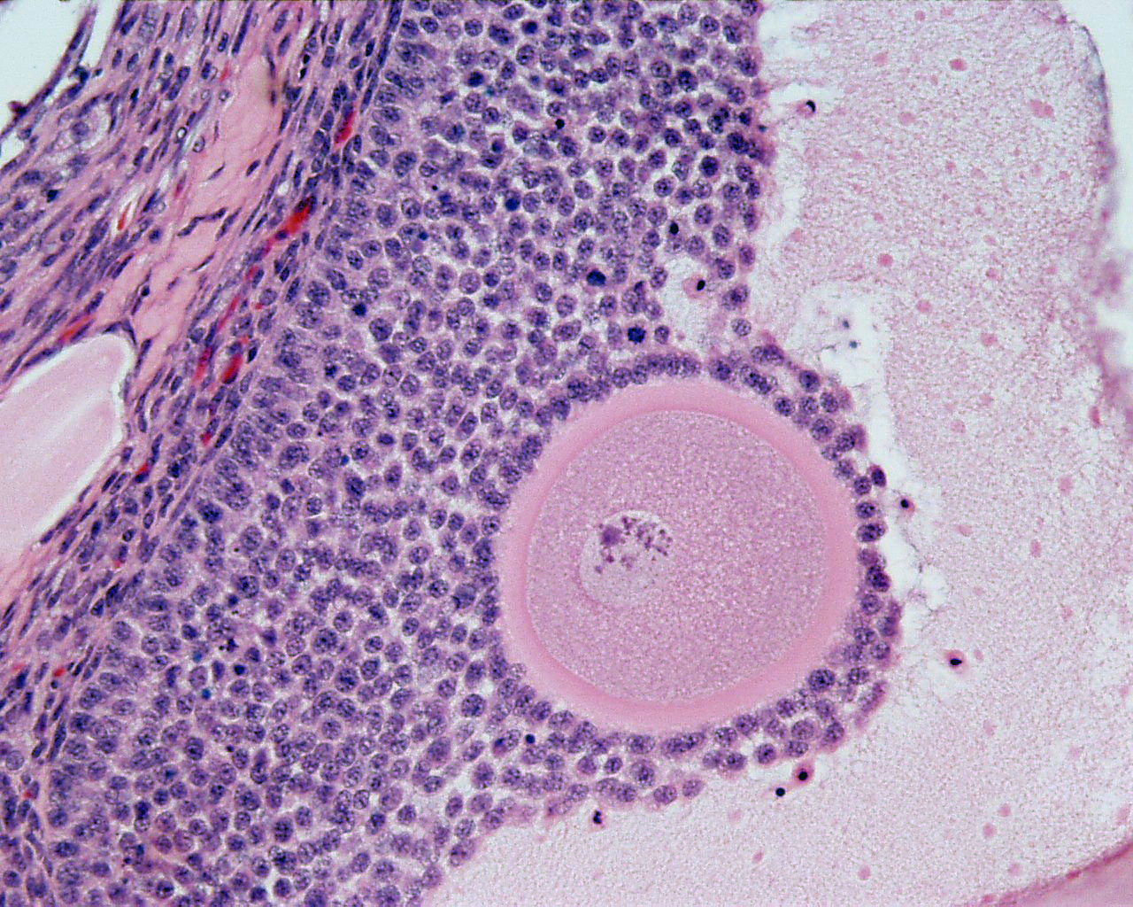
Ovary - Antral Follicle
ovary, monkey (Stain - Haematoxylin Eosin)
- Small fluid-filled spaces become visible between the granulosa cells as the follicle reaches a diameter of about 400 µm.
- These spaces enlarge and fuse to form the follicular antrum, which is the defining feature of the secondary follicle.
- The oocyte is now located eccentric in the follicle in the cumulus oophorus, where it is surrounded by granulosa cells.
- The theca folliculi differentiates with the continued growth of the follicle into a theca interna and a theca externa.
- Vascularization of the theca interna improves, and the spindle-shaped or polyhedral cells in this layer start to produce oestrogens.
- The theca externa retains the characteristics of a highly cellular connective tissue with smooth muscle cells.
- The oocyte of the secondary follicle reaches a diameter of about 125 µm.
- The follicle itself reaches a diameter of about 10-15 mm.
- Links: Image-Secondary Follicle labeled | Image-Secondary Follicle labeled | Ovary Development | Oocyte Development
Ovary histology: Tunica Albuginea x20 | Tunica albuginea, Germinal epithelium x40 |
Primary follicle, primordial follicle, oocyte, x40 | Secondary follicle, cumulus oophorus, zona pelucida, granulosa cells, oocyte x20 | Corpus luteum, theca lutein cells, granulosa lutein cells, Loupe | Corpus luteum, theca lutein cells, granulosa lutein cells, x10 | Corpus luteum, theca lutein cells, granulosa lutein cells, x40 | Corpus albicans, primary follicle, primordial follicle, granulosa cells, oocyte x20 | Menstrual Cycle | Ovary Development
Links: Histology | Histology Stains | Blue Histology images copyright Lutz Slomianka 1998-2009. The literary and artistic works on the original Blue Histology website may be reproduced, adapted, published and distributed for non-commercial purposes. See also the page Histology Stains.
Cite this page: Hill, M.A. (2024, April 19) Embryology Ovary histology 006.jpg. Retrieved from https://embryology.med.unsw.edu.au/embryology/index.php/File:Ovary_histology_006.jpg
- © Dr Mark Hill 2024, UNSW Embryology ISBN: 978 0 7334 2609 4 - UNSW CRICOS Provider Code No. 00098G
Cite this page: Hill, M.A. (2024, April 19) Embryology Ovary histology 006.jpg. Retrieved from https://embryology.med.unsw.edu.au/embryology/index.php/File:Ovary_histology_006.jpg
- © Dr Mark Hill 2024, UNSW Embryology ISBN: 978 0 7334 2609 4 - UNSW CRICOS Provider Code No. 00098G
File history
Click on a date/time to view the file as it appeared at that time.
| Date/Time | Thumbnail | Dimensions | User | Comment | |
|---|---|---|---|---|---|
| current | 21:35, 5 May 2011 | | 1,280 × 1,024 (424 KB) | S8600021 (talk | contribs) | |
| 21:11, 23 February 2011 | 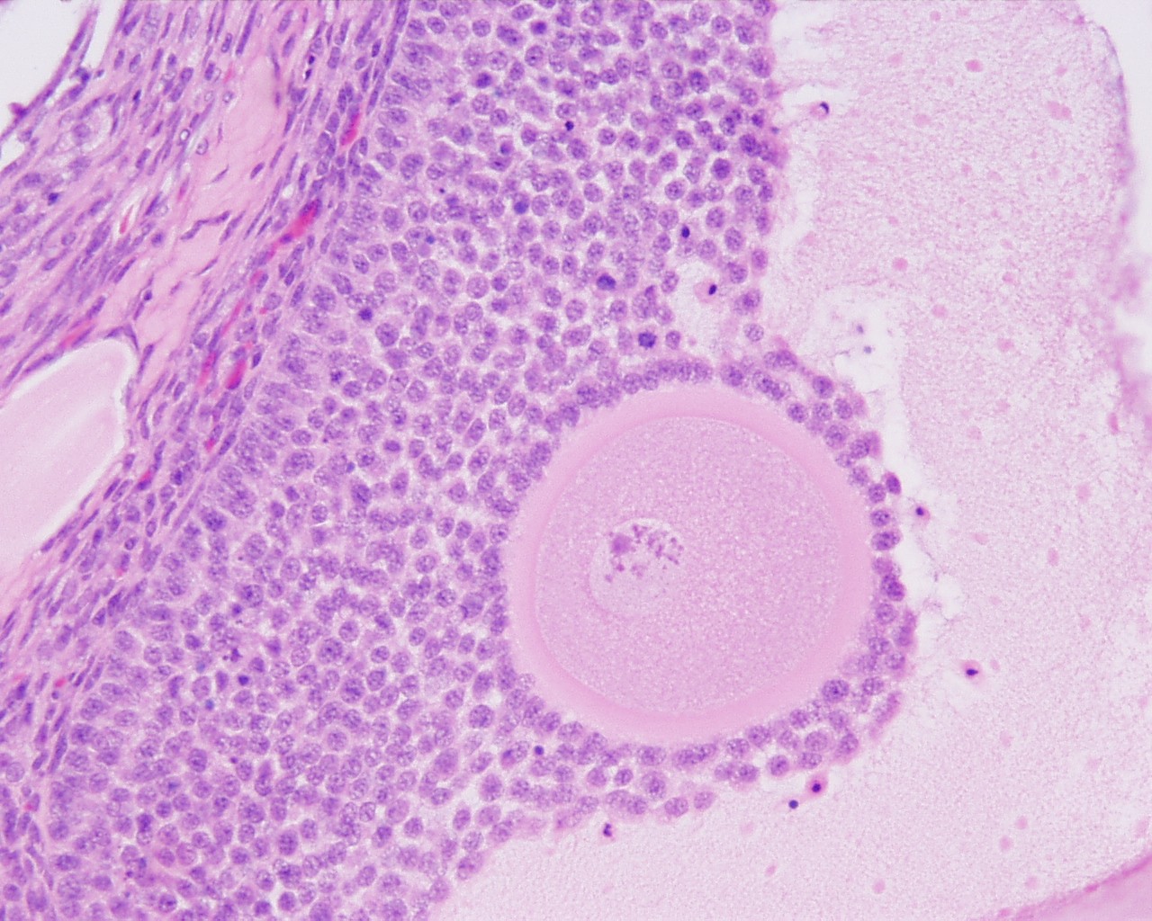 | 1,280 × 1,024 (340 KB) | S8600021 (talk | contribs) | File:Ovary_histology_006.jpg Ovary,_monkey_H&E_reproductive_system,_female,_secondary_follicle,_cumulus_oophorus,_zona_pelucida,_granulosa_cells,_oocyte_x20.jpg {{Ovary Histology}} {{Template:Blue Histology}} Category:Monkey Category:Genital |
You cannot overwrite this file.
File usage
The following 6 pages use this file: