File:Ossification centre.jpg
From Embryology
No higher resolution available.
Ossification_centre.jpg (450 × 600 pixels, file size: 101 KB, MIME type: image/jpeg)
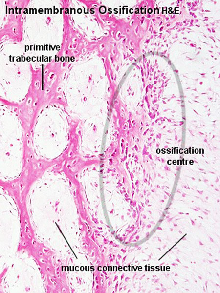
Intramembranous Ossification Centre
- Histological areas of a gradual transition from connective tissue to bone.
- Light, pinkish bone matrix is found between the osteoblasts.
- Depending on the state of development of the bone, it is occasionally possible to find bone trabeculae which are lined by a layer of osteoblasts.
- These osteblasts are depositing the first lamellae on the already existing trabeculae.
- The trabeculae will therefore have a core of woven bone, which is surrounded by lamellar bone.
- Compare the shapes, sizes and frequencies of lacunae in lamellar and woven bone if both types of bone are present.
Links: Histology | Histology Stains | Blue Histology images copyright Lutz Slomianka 1998-2009. The literary and artistic works on the original Blue Histology website may be reproduced, adapted, published and distributed for non-commercial purposes. See also the page Histology Stains.
Cite this page: Hill, M.A. (2024, April 20) Embryology Ossification centre.jpg. Retrieved from https://embryology.med.unsw.edu.au/embryology/index.php/File:Ossification_centre.jpg
- © Dr Mark Hill 2024, UNSW Embryology ISBN: 978 0 7334 2609 4 - UNSW CRICOS Provider Code No. 00098G
Original Image name: Imos10he.jpg
File history
Click on a date/time to view the file as it appeared at that time.
| Date/Time | Thumbnail | Dimensions | User | Comment | |
|---|---|---|---|---|---|
| current | 15:57, 13 May 2012 | | 450 × 600 (101 KB) | Z8600021 (talk | contribs) | |
| 11:30, 11 September 2009 |  | 300 × 400 (66 KB) | S8600021 (talk | contribs) | Ossification centre Histological areas of a gradual transition from connective tissue to bone. Light, pinkish bone matrix is found between the osteoblasts. Depending on the state of development of the bone, it is occasionally possible to find bone trabec |
You cannot overwrite this file.
File usage
The following 14 pages use this file:
- 2009 Lecture 13
- 2010 BGD Lecture - Development of the Embryo/Fetus 2
- 2010 BGD Practical 6 - Week 7
- 2010 Lecture 13
- ANAT2241 Bone, Bone Formation and Joints
- ANAT2341 Lab 6 - Fetal
- BGDA Lecture - Development of the Embryo/Fetus 2
- BGDA Practical 7 - Week 7
- BGDB Face and Ear - Fetal
- Bone Development
- Bone Histology
- Lecture - Fetal Development
- Lecture - Musculoskeletal Development
- Musculoskeletal System - Bone Development