File:Multilobed placenta MRI01.jpg
Multilobed_placenta_MRI01.jpg (600 × 599 pixels, file size: 145 KB, MIME type: image/jpeg)
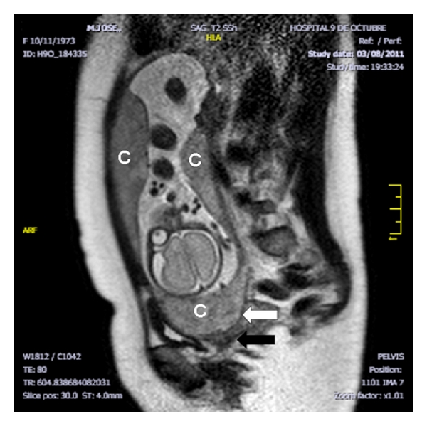
multilobed placenta MRI
GA week 27 magnetic resonance image.
- multilobed placenta succenturiata
- succenturiata - an accessory portion attached to the main placenta by an artery or vein.
- Placenta succenturiata is a type of multilobed placenta in which one or various cotyledons are separated from the main lobe by membranous areas.
- umbilical cord clearly inserted into this cervical anomalous cotyledon
- c - three placental portions
- white arrow - placental portion situated below the fetal head covering the internal os
- black arrow - very short and incompetent cervix
Reference
<pubmed>22481947</pubmed>| PMC3299365
Copyright
© 2012 José Morales-Roselló and Núria Peralta Llorens. This is an open access article distributed under the Creative Commons Attribution License, which permits unrestricted use, distribution, and reproduction in any medium, provided the original work is properly cited.
CRIM2012-293156.002.jpg
Cite this page: Hill, M.A. (2024, April 18) Embryology Multilobed placenta MRI01.jpg. Retrieved from https://embryology.med.unsw.edu.au/embryology/index.php/File:Multilobed_placenta_MRI01.jpg
- © Dr Mark Hill 2024, UNSW Embryology ISBN: 978 0 7334 2609 4 - UNSW CRICOS Provider Code No. 00098G
File history
Click on a date/time to view the file as it appeared at that time.
| Date/Time | Thumbnail | Dimensions | User | Comment | |
|---|---|---|---|---|---|
| current | 20:31, 11 June 2013 | | 600 × 599 (145 KB) | Z8600021 (talk | contribs) | ==multilobed placenta MRI== * multilobed placenta succenturiata * Placenta succenturiata is a type of multilobed placenta in which one or various cotyledons are separated from the main lobe by membranous areas. * umbilical cord clearly inserted into ... |
You cannot overwrite this file.