File:Mouse zygote pronuclei 02.jpg

Original file (1,000 × 501 pixels, file size: 84 KB, MIME type: image/jpeg)
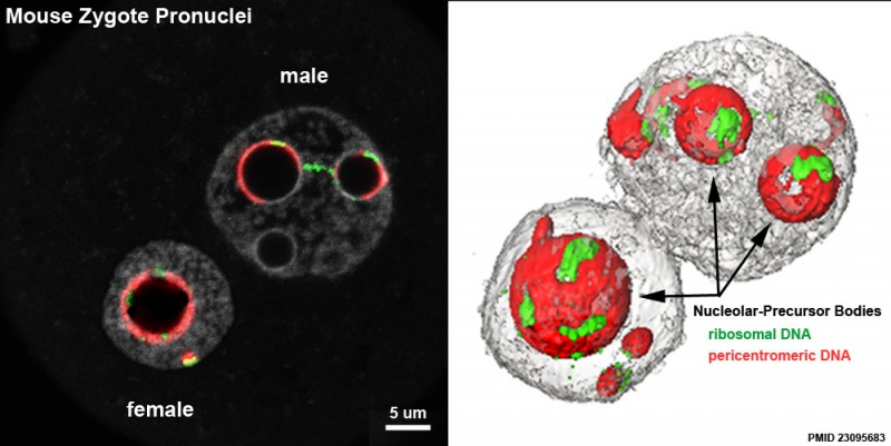
Mouse Zygote Pronuclei
Distribution of the pericentromeres, centromeres, and rDNA FISH signals in late 1-cell stage embryos (fluorescent and 3D reconstruction). Transcription of ribosomal DNA (rDNA) is switched off in early mouse embryos and nucleoli are not present.
Most rDNA signals are around the Nucleolar-Precursor Bodies (NPBs), though some signals associated with pericentromeric filaments (extending from the NPBs towards the nuclear periphery) as well as rDNA signals joining two NPBs.
1-cell embryos at the PN4 stage (fertilization occurred at about 12 after hCG injection (hphCG); zygote collected at 27 hours)
- red - pericentromeric probes
- green - ribosomal DNA probes
- grey - DNA counterstained with Yopro-1
Bar = 5 μm.
Embryonic gene expression (EGA, embryonic genome activation) is a rapid increase in the synthesis of transcripts. The reinitiation of rDNA transcription occurs at the end of the 2-cell stage, at the surface of the NPBs.
Fertilization occurred at about 12 hours after hCG injection, which was used as the reference point for embryonic development (hours post-hCG i.e. hphCG).
Reference
<pubmed>23095683</pubmed>| BMC Dev Biol.
Copyright
© 2012 Aguirre Lavin et al.; licensee BioMed Central Ltd. This is an Open Access article distributed under the terms of the Creative Commons Attribution License ( http://creativecommons.org/licenses/by/2.0), which permits unrestricted use, distribution, and reproduction in any medium, provided the original work is properly cited.
Figure 1. Panel E and F cropped, resized and relabelled. Text modified from paper and legend.
File history
Click on a date/time to view the file as it appeared at that time.
| Date/Time | Thumbnail | Dimensions | User | Comment | |
|---|---|---|---|---|---|
| current | 10:43, 29 December 2012 | | 1,000 × 501 (84 KB) | Z8600021 (talk | contribs) |
You cannot overwrite this file.
File usage
The following page uses this file: